Japanese
English
- 有料閲覧
- Abstract 文献概要
- 1ページ目 Look Inside
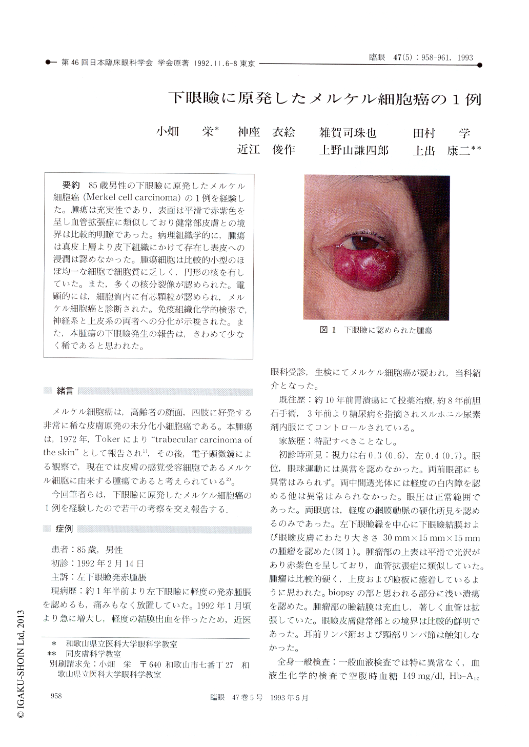
85歳男性の下眼瞼に原発したメルケル細胞癌(Merkel cell carcinoma)の1例を経験した。腫瘍は充実性であり,表面は平滑で赤紫色を呈し血管拡張症に類似しており健常部皮膚との境界は比較的明瞭であった。病理組織学的に,腫瘍は真皮上層より皮下組織にかけて存在し表皮への浸潤は認めなかった。腫瘍細胞は比較的小型のほぼ均一な細胞で細胞質に乏しく,円形の核を有していた。また,多くの核分裂像が認められた。電顕的には,細胞質内に有芯顆粒が認められ,メルケル細胞癌と診断された。免疫組織化学的検索で,神経系と上皮系の両者への分化が示唆された。また,本腫瘍の下眼瞼発生の報告は,きわめて少なく稀であると思われた。
A 85-year-old male was diagnosed as Merkel cellcarcinoma in the left lower eyelid. The tumor wasa cutaneous nodule with reddish-purple overlyingskin. It was well-defined and resembled an an-giomatous lesion. Histologically, the tumor invadedthe dermis and subcutaneous tissue of the eyelidand did not extend into the epidermis. The tumorcells were small-sized and showed a monotonouslyuniform appearance. They had round nuclei andscant cytoplasm. Mitotic activity was high. Elec-tron microscopy showed membrane-bounded densecore granules in the cytoplasm which were specificto Merkel cells. Immunohistochemically, the tumorwas positively stained for neuron-specific enolase,chromogranin, cytokeratin and epithelial mem-brane antigen. These features showed neural andepidermal differentiation of tumor cells. To ourknowledge, only 3 cases of Merkel cell carcinomaof the lower eyelid have been reported so far.

Copyright © 1993, Igaku-Shoin Ltd. All rights reserved.


