Japanese
English
- 有料閲覧
- Abstract 文献概要
- 1ページ目 Look Inside
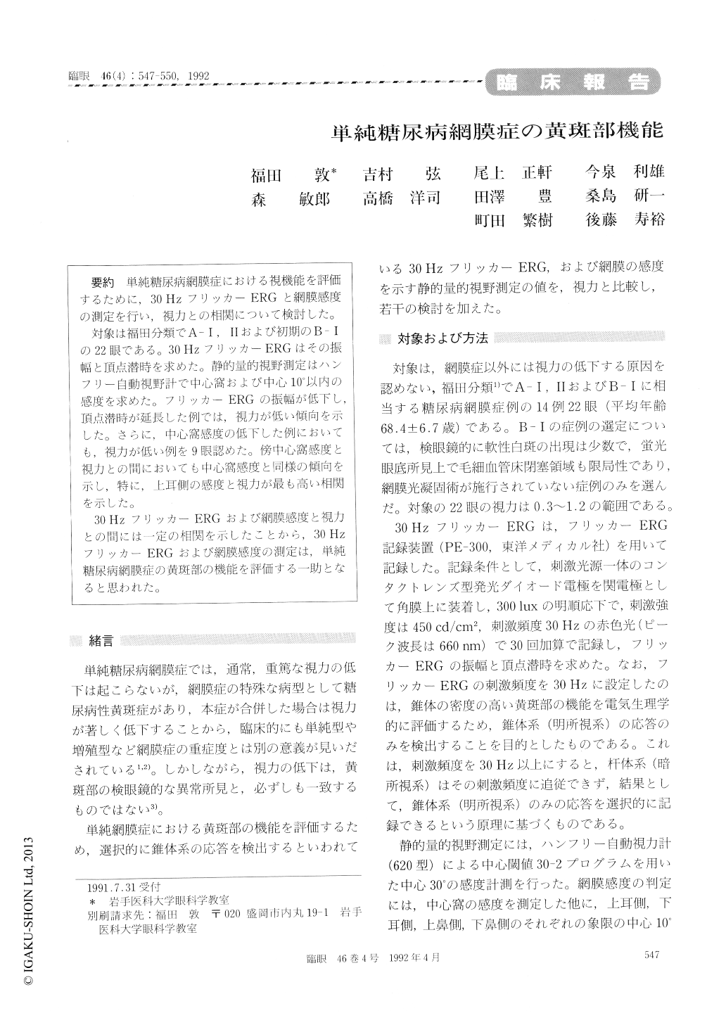
単純糖尿病網膜症における視機能を評価するために,30HzフリッカーERGと網膜感度の測定を行い,視力との相関について検討した。
対象は福田分類でA—Ⅰ,Ⅱおよび初期のB—Ⅰの22眼である。30HzフリッカーERGはその振幅と頂点潜時を求めた。静的量的視野測定はハンフリー自動視野計で中心窩および中心10°以内の感度を求めた。フリッカーERGの振幅が低下し,頂点潜時が延長した例では,視力が低い傾向を示した。さらに,中心窩感度の低下した例においても,視力が低い例を9眼認めた。傍中心窩感度と視力との間においても中心窩感度と同様の傾向を示し,特に,上耳側の感度と視力が最も高い相関を示した。
30HzフリッカーERGおよび網膜感度と視力との間には一定の相関を示したことから,30HzフリッカーERGおよび網膜感度の測定は,単純糖尿病網膜症の黄斑部の機能を評価する一助となると思われた。
We evaluated 22 eyes in 14 patients with simple diabetic retinopathy regarding visual acuity, flicker electroretinogram (ERG) and static retinal sensi-tivity. All the eyes were in the early stage of retinopathy graded as A-Ⅰ, A-Ⅱ or B-Ⅰaccord-ing to Fukuda's classification. We measured the amplitudes and latencies of 30 Hz flicker ERG. We also measured the foveal sensitivity and mean sen-sitivity in each of the 4 quadrants within 10 degrees from the fixation point. Static visual field was measured by program C-30-2 of Humphrey field analyzer. The amplitudes of flicker ERG were depressed in eyes with low visual acuity. The peak latency of flicker ERG was prolonged parallel to decrease in visual acuity. Foveal sensitivity de-creased parallel to visual acuity. Retinal sensitivity and visual acuity showed highest correlation in the parafoveal superotemporal quadrant. The findings suggest that changes in amplitude and peak latency in flicker ERG as well as retinal sensitivity are early indicators of impaired visual function in early diabetic maculopathy.

Copyright © 1992, Igaku-Shoin Ltd. All rights reserved.


