Japanese
English
- 有料閲覧
- Abstract 文献概要
- 1ページ目 Look Inside
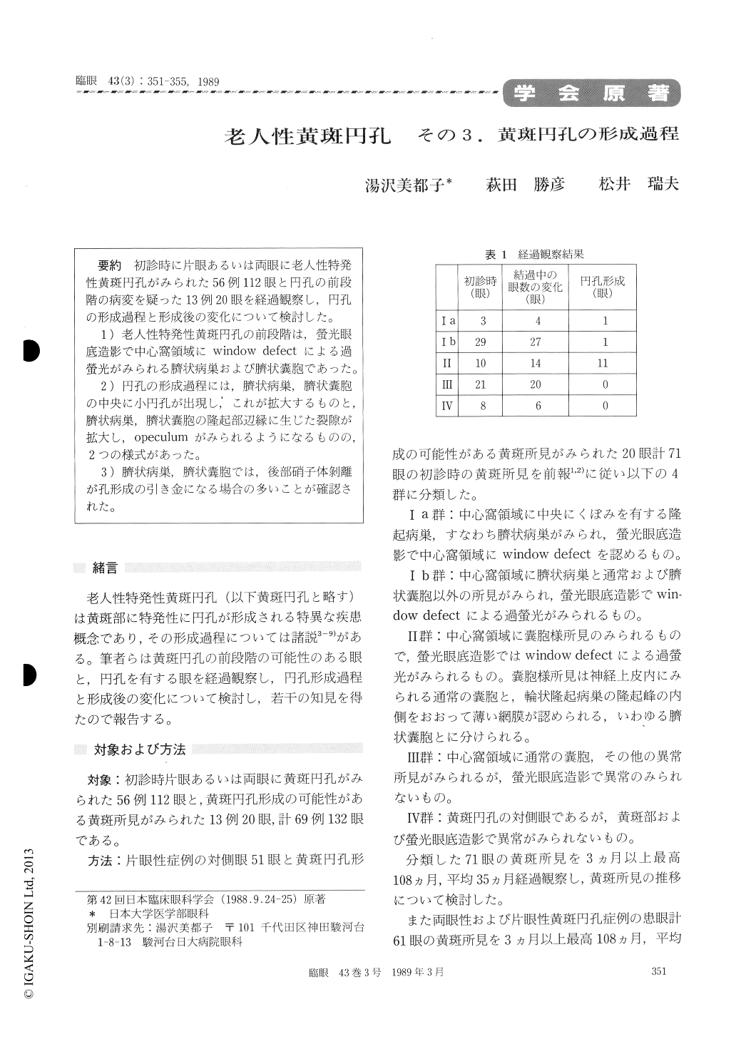
初診時に片眼あるいは両眼に老人性特発性黄斑円孔がみられた56例112眼と円孔の前段階の病変を疑った13例20眼を経過観察し,円孔の形成過程と形成後の変化について検討した。
1)老人性特発性黄斑円孔の前段階は,螢光眼底造影で中心窩領域にwindow defectによる過螢光がみられる臍状病巣および臍状嚢胞であった。
2)円孔の形成過程には,臍状病巣,臍状嚢胞の中央に小円孔が出現し,これが拡大するものと,臍状病巣,臍状嚢胞の隆起部辺縁に生じた裂隙が拡大し,opeculumがみられるようになるものの,2つの様式があった。
3)臍状病巣,臍状嚢胞では,後部硝子体剥離が孔形成の引き金になる場合の多いことが確認された。
We evaluated a series of 112 eyes in 56 patients with unilateral or bilateral idiopathic senile macular hole. We also evaluated another series of 20 eyes in 13 patients which manifested impending or evolving macular hole formation.
We could identify two pathological features which developed into manifest macular hole. The first was a small hole in the center of navel-like lesion or navel cyst in the macula. The small hole gradually enlarged to form manifest macular hole. This was seen in 4 eyes. The second was a tear which developed along the margin of the navel-like lesion or navel cyst. The tear later enlarged to form an operculum. This finding was confirmed in 6 eyes. The navel-like lesion appeared as a circular eleva-tion surrounding the fovea. The navel cyst showed a circular elevated lesion covered with a thin layer of the sensory retina.
Posterior vitreous separation in the foveal area contributed to the formation of macular hole in 8 eyes.

Copyright © 1989, Igaku-Shoin Ltd. All rights reserved.


