Japanese
English
- 有料閲覧
- Abstract 文献概要
- 1ページ目 Look Inside
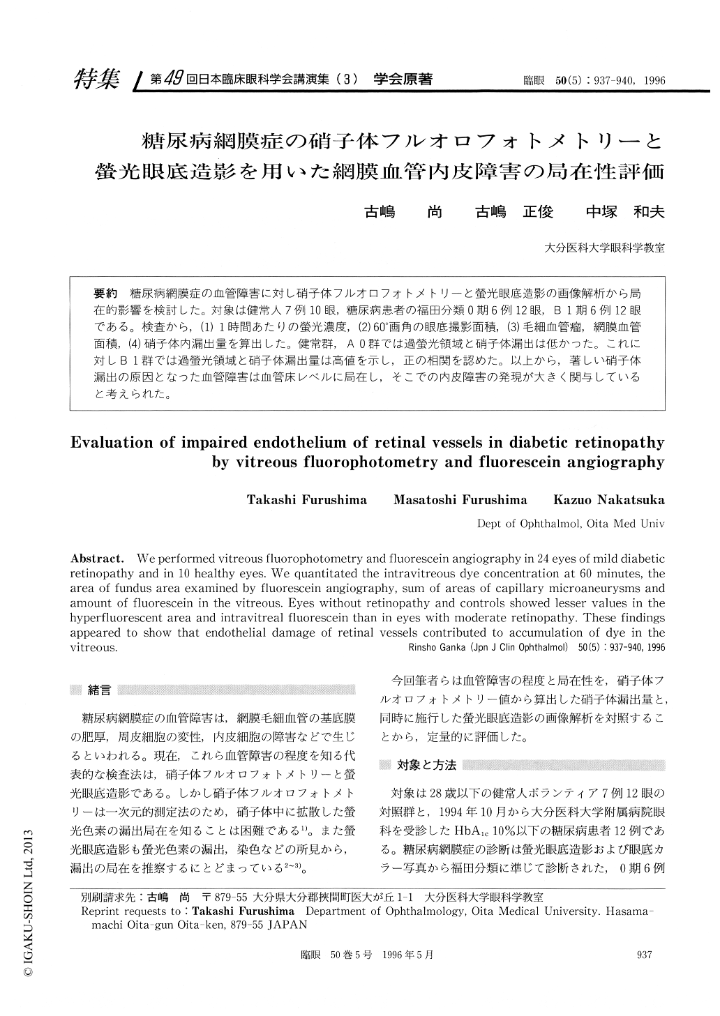
糖尿病網膜症の血管障害に対し硝子体フルオロフォトメトリーと螢光眼底造影の画像解析から局在的影響を検討した。対象は健常人7例10眼,糖尿病患者の福田分類0期6例12眼,B1期6例12眼である。検査から,(1)1時間あたりの螢光濃度,(2)60°画角の眼底撮影面積,(3)毛細血管瘤,網膜血管面積,(4)硝子体内漏出量を算出した。健常群,AO群では過螢光領域と硝子体漏出は低かった。これに対しB1群では過螢光領域と硝子体漏出量は高値を示し,正の相関を認めた。以上から,著しい硝子体漏出の原因となった血管障害は血管床レベルに局在し,そこでの内皮障害の発現が大きく関与していると考えられた。
We performed vitreous fluorophotometry and fluorescein angiography in 24 eyes of mild diabetic retinopathy and in 10 healthy eyes. We quantitated the intravitreous dye concentration at 60 minutes, the area of fundus area examined by fluorescein angiography, sum of areas of capillary microaneurysms and amount of fluorescein in the vitreous. Eyes without retinopathy and controls showed lesser values in the hyperfluorescent area and intravitreal fluorescein than in eyes with moderate retinopathy. These findings appeared to show that endothelial damage of retinal vessels contributed to accumulation of dye in the vitreous.

Copyright © 1996, Igaku-Shoin Ltd. All rights reserved.


