Japanese
English
- 有料閲覧
- Abstract 文献概要
- 1ページ目 Look Inside
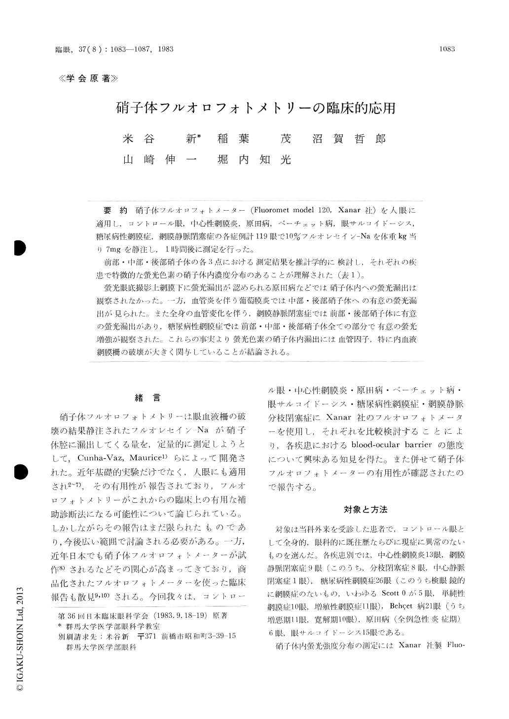
硝子体フルオロフォトメーター(Fluoromet model 120, Xanar社)を人眼に適用し,コントロール眼,中心性網膜炎,原田病,ベーチェット病,眼サルコイドーシス,糖尿病性網膜症,網膜静脈閉塞症の各症例計119眼で10%フルオレセイン—Naを体重kg当り7mgを静注し,1時間後に測定を行った。
前部・中部・後部硝子体の各3点における測定結果を推計学的に検討し,それぞれの疾患で特徴的な螢光色素の硝子体内濃度分布のあることが理解された(表1)。
螢光眼底撮影上網膜下に螢光漏出が認められる原田病などでは硝子体内への螢光漏出は観察されなかった。一方,血管炎を伴う葡萄膜炎では中部・後部硝子体への有意の螢光漏出が見られた。また全身の血管変化を伴う,網膜静脈閉塞症では前部・後部硝子体に有意の螢光漏出があり,糖尿病性網膜症では前部・中部・後部硝子体全ての部分で有意の螢光増強が観察された。これらの事実より螢光色素の硝子体内漏出には孟1管因子,特に内血液網膜柵の破壊が大きく関与していることが結論される。
We performed vitreous fluorophotometry in a total of 119 eyes with central serous retinopathy, diabetic retinopathy, branch retinal vein occlusion or uveitis using a high-sensitive vitreous fluoropho-tometer Fluoromet model 120 (Xanar). 29 normal eyes served as control. Detailed fundus examina-tions and fluorescein angiography were performed prior to fluorophotometry. Concentration profiles were recorded for 60 minutes following i.v. injec-tion of 10% fluorescein sodium (7mg/kg). The data were processed at three cardinal points in the vitre-ous cavity, i.e. anterior, middle and posterior vitreous.

Copyright © 1983, Igaku-Shoin Ltd. All rights reserved.


