Japanese
English
- 有料閲覧
- Abstract 文献概要
- 1ページ目 Look Inside
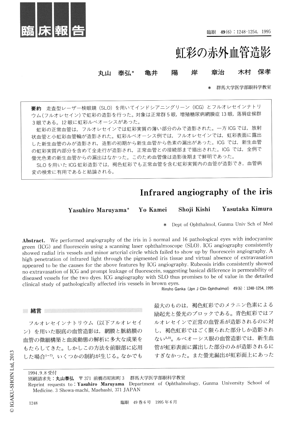
走査型レーザー検眼鏡(SLO)を用いてインドシアニングリーン(ICG)とフルオレセインナトリウム(フルオレセイン)で虹彩の造影を行った。対象は正常群5眼,増殖糖尿病網膜症13眼,落屑症候群3眼である。12眼に虹彩ルベオーシスがあった。
虹彩の正常血管は,フルオレセインでは虹彩実質の薄い部分のみで造影された。一方ICGでは,放射状血管と小虹彩血管輪が造影された。虹彩ルベオーシス例では,フルオレセインでは,虹彩表面に露出した新生血管のみが造影され,造影の初期から新生血管から色素の漏出があった。ICGでは,新生血管の虹彩実質内部分を含めて全走行が造影され,正常血管との接続部まで描出された。ICGでは,全例で螢光色素の新生血管からの漏出はなかった。このため血管像は造影後期まで鮮明であった。
SLOを用いたICG虹彩造影では,褐色虹彩でも正常血管を含む虹彩実質内の血管が造影でき,血管病変の検索に有用であると結論される。
We performed angiography of the iris in 5 normal and 16 pathological eyes with indocyanine green (ICG) and fluorescein using a scanning laser ophthalmoscope (SLO). ICG angiography consistently showed radial iris vessels and minor arterial circle which failed to show up by fluorescein angiography. A high penetration of infrared light through the pigmented iris tissue and virtual absence of extravasation appeared to be the causes for the above features by ICG angiography. Rubeosis iridis consistently showed no extravasation of ICG and prompt leakage of fluorescein, suggesting basical difference in permeability of diseased vessels for the two dyes. ICG angiography with SLO thus promises to be of value in the detailed clinical study of pathologically affected iris vessels in brown eyes.

Copyright © 1995, Igaku-Shoin Ltd. All rights reserved.


