Japanese
English
- 有料閲覧
PPV購入案内
1,320円 (1,200円+税10%) こちらは、1527ページから1534ページの文献です。購入には医書.jp本会員登録が必要です。
会員登録完了後に改めて上記「購入サイトへ」を選択してください。
または「購入サイトへ」選択後に「購入ログイン」、「新規会員登録」の順に進んでください。
会員登録について詳しくはをご確認ください。
- Abstract 文献概要
- 1ページ目 Look Inside
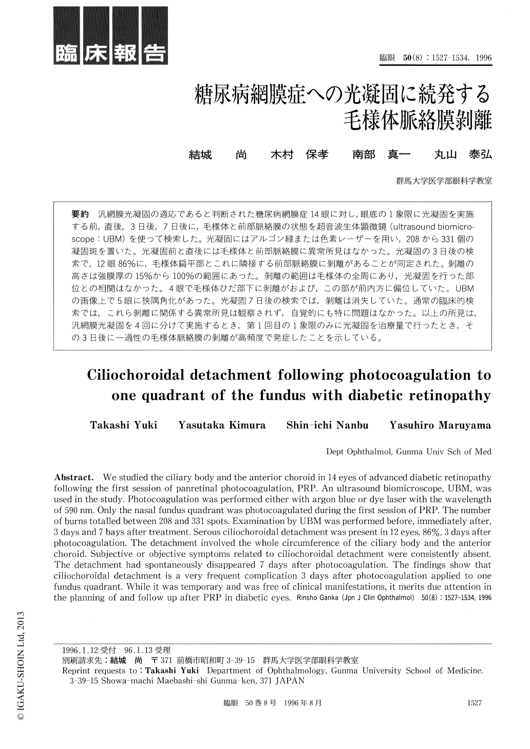
汎網膜光凝固の適応であると判断された糖尿病網膜症14眼に対し,眼底の1象限に光凝固を実施する前,直後,3日後,7日後に,毛様体と前部脈絡膜の状態を超音波生体顕微鏡(ultrasound biomicro—scope:UBM)を使って検索した。光凝固にはアルゴン緑または色素レーザーを用い,208から331個の凝固斑を置いた。光凝固前と直後には毛様体と前部脈絡膜に異常所見はなかった。光凝固の3日後の検索で,12眼86%に,毛様体扁平部とこれに隣接する前部脈絡膜に剥離があることが同定された。剥離の高さは強膜厚の15%から100%の範囲にあった。剥離の範囲は毛様体の全周にあり,光凝固を行った部位との相関はなかった。4眼で毛様体ひだ部下に剥離がおよび,この部が前内方に偏位していた。UBMの画像上で5眼に狭隅角化があった。光凝固7日後の検索では,剥離は消失していた。通常の臨床的検索では,これら剥離に関係する異常所見は観察されず,自覚的にも特に問題はなかった。以上の所見は,汎網膜光凝固を4回に分けて実施するとき,第1回目の1象限のみに光凝固を治療量で行ったとき,その3日後に一過性の毛様体脈絡膜の剥離が高頻度で発症したことを示している。
We studied the ciliary body and the anterior choroid in 14 eyes of advanced diabetic retinopathy following the first session of panretinal photocoagulation, PRP. An ultrasound biomicroscope, UBM, was used in the study. Photocoagulation was performed either with argon blue or dye laser with the wavelength of 590nm. Only the nasal fundus quadrant was photocoagulated during the first session of PRP. The number of burns totalled between 208 and 331 spots. Examination by UBM was performed before, immediately after,3 days and 7 bays after treatment. Serous ciliochoroidal detachment was present in 12 eyes, 86%, 3 days after photocoagulation. The detachment involved the whole circumference of the ciliary body and the anterior choroid. Subjective or objective symptoms related to ciliochoroidal detachment were consistently absent.The detachment had spontaneously disappeared 7 days after photocoagulation. The findings show that ciliochoroidal detachment is a very frequent complication 3 days after photocoagulation applied to one fundus quadrant. While it was temporary and was free of clinical manifestations, it merits due attention in the planning of and follow up after PRP in diabetic eyes.
Copyright © 1996, Igaku-Shoin Ltd. All rights reserved.