Japanese
English
- 有料閲覧
- Abstract 文献概要
- 1ページ目 Look Inside
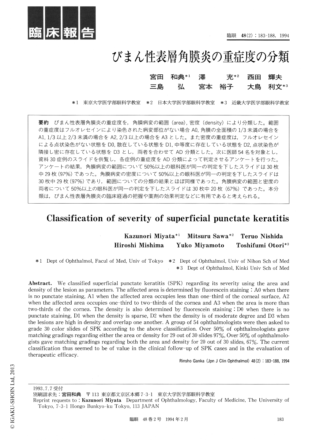
びまん性表層角膜炎の重症度を,角膜病変の範囲(area),密度(density)により分類した。範囲の重症度はフルオレセインにより染色された病変部位がない場合A0,角膜の全面積の1/3未満の場合をA1,1/3以上2/3未満の場合をA2,2/3以上の場合をA3とした。また密度の重症度は,フルオレセインによる点状染色がない状態をD0,散在している状態をD1,中等度に存在している状態をD2,点状染色が隣接し密に存在している状態をD3とし,両者を合わせてAD分類とした。次に医師54名を対象とし,資料30症例のスライドを供覧し,各症例の重症度をAD分類によって判定させるアンケートを行った。アンケートの結果,角膜病変の範囲について50%以上の眼科医が同一の判定を下したスライドは30枚中29枚(97%)であった。角膜病変の密度について50%以上の眼科医が同一の判定を下したスライドは30枚中29枚(97%)であり,範囲についての分類の結果とほぼ同様であった。角膜病変の範囲と密度の両者について50%以上の眼科医が同一の判定を下したスライドは30枚中20枚(67%)であった。本分類は,びまん性表層角膜炎の臨床経過の把握や薬剤の効果判定などに有用であると考えられる。
We classified superficial punctate keratitis (SPK) regarding its severity using the area and density of the lesion as parameters. The affected area is determined by fluorescein staining: A0 when there is no punctate staining, A1 when the affected area occupies less than one-third of the corneal surface, A2 when the affected area occupies one-third to two-thirds of the cornea and A3 when the area is more than two-thirds of the cornea. The density is also determined by fluorescein staining :D0 when there is no punctate staining, Dl when the density is sparse, D2 when the density is of moderate degree and D3 when the lesions are high in density and overlap one another. A group of 54 ophthalmologists were then asked to grade 30 color slides of SPK according to the above classification. Over 50% of ophthalmologists gave matching gradings regarding either the area or density for 29 out of 30 slides 97%, Over 50% of ophthalmolo-gists gave matching gradings regarding both the area and density for 20 out of 30 slides, 67%. The current classification thus seemed to be of value in the clinical follow-up of SPK cases and in the evaluation of therapeutic efficacy.

Copyright © 1994, Igaku-Shoin Ltd. All rights reserved.


