Japanese
English
- 有料閲覧
- Abstract 文献概要
- 1ページ目 Look Inside
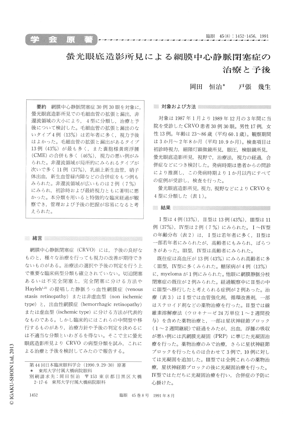
網膜中心静脈閉塞症30例30眼を対象に螢光眼底造影所見での毛細血管の拡張と漏出,非還流領域の大小により,4型に分類し,治療と予後について検討した。毛細血管の拡張と漏出のないタイプ4例(13%)は若年者に多く,視力予後はよかった。毛細血管の拡張と漏出があるタイプ13例(43%)が最も多く,また嚢胞様黄斑浮腫(CME)の合併も多く(46%),視力の悪い例がみられた。非還流領域が局所的にみられるタイプが次いで多く11例(37%),乳頭上新生血管,硝子体出血,新生血管緑内障などの合併症をもつ例もみられた。非還流領域が広いものは2例(7%)にみられ,初診時および最終視力ともに著明に悪かった。本分類を用いると特徴的な臨床経過が観察でき,管理および予後の把握が容易になると考えられた。
We evaluated the clinical feature of 30 cases (30 eyes) who had central retinal vein occlusion. According to the fluorescein angiography, these cases were classified into the following four cate-gories of retinal capillary perfusion:
1) Minimal capillary dilatation and leakage (13%)-cases which belonged to mainly young patients, and led to good visual prog-nosis.
2) Marked capillary dilatation and leakage (43%) cases which had complications ofcystoid macular edema (46%) and some of them showed poor visual acuity.
3) Partial non perfusion area (37%)-cases which included optic disc neovascularization, vitreous hemorrhage and neovascular glau-coma.
4) Wide non perfusion area (7%)-cases which had remarkably poor visual prognosis.
Thus, this classification provided characteritic clinical features, and it led to management and prognosis easily.

Copyright © 1991, Igaku-Shoin Ltd. All rights reserved.


