Japanese
English
- 有料閲覧
- Abstract 文献概要
- 1ページ目 Look Inside
- 参考文献 Reference
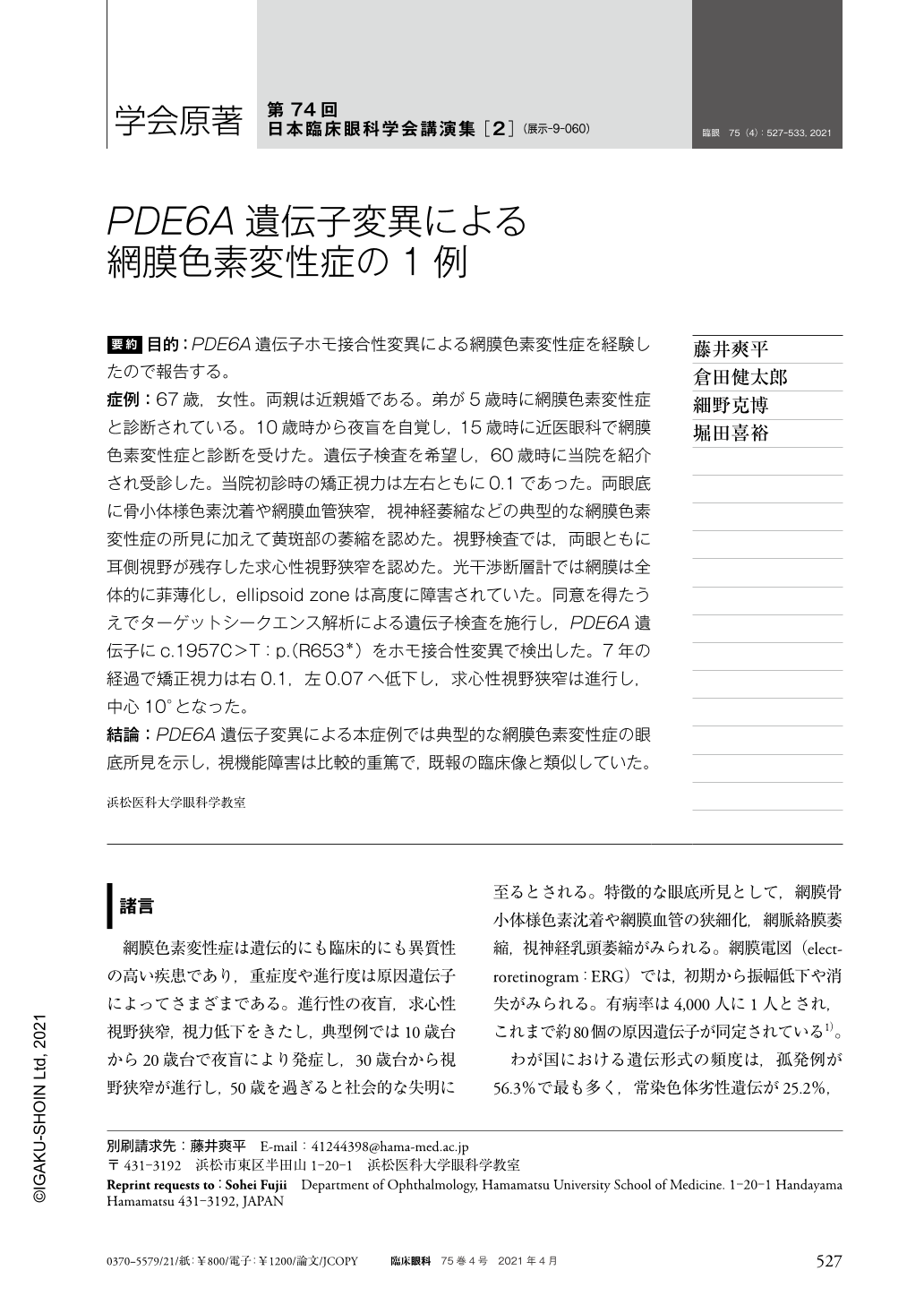
要約 目的:PDE6A遺伝子ホモ接合性変異による網膜色素変性症を経験したので報告する。
症例:67歳,女性。両親は近親婚である。弟が5歳時に網膜色素変性症と診断されている。10歳時から夜盲を自覚し,15歳時に近医眼科で網膜色素変性症と診断を受けた。遺伝子検査を希望し,60歳時に当院を紹介され受診した。当院初診時の矯正視力は左右ともに0.1であった。両眼底に骨小体様色素沈着や網膜血管狭窄,視神経萎縮などの典型的な網膜色素変性症の所見に加えて黄斑部の萎縮を認めた。視野検査では,両眼ともに耳側視野が残存した求心性視野狭窄を認めた。光干渉断層計では網膜は全体的に菲薄化し,ellipsoid zoneは高度に障害されていた。同意を得たうえでターゲットシークエンス解析による遺伝子検査を施行し,PDE6A遺伝子にc.1957C>T:p.(R653*)をホモ接合性変異で検出した。7年の経過で矯正視力は右0.1,左0.07へ低下し,求心性視野狭窄は進行し,中心10°となった。
結論:PDE6A遺伝子変異による本症例では典型的な網膜色素変性症の眼底所見を示し,視機能障害は比較的重篤で,既報の臨床像と類似していた。
Abstract Purpose:We describe a case of retinitis pigmentosa(RP)caused by a single homozygous PDE6A mutations.
Case:The patient was a 67-year-old woman born to consanguineous parents. Her brother was diagnosed with RP at 5 years of age. She developed night blindness at 10 years of age and was diagnosed with RP at 15 years of age by a local doctor. She was referred to our hospital at 60 years of age. Her best-corrected visual acuity(BCVA)was 0.1 in both eyes, and fundoscopy revealed bone spicule pigmentation, narrowed retinal vessels, waxy pallor of the optic discs, and macular atrophy. Goldmann perimetry revealed concentric constriction with a residual temporal island. Optical coherence tomography revealed thinned retinal layers and a disrupted ellipsoid zone. Genetic analysis revealed a single homozygous PDE6A mutations in the patient. Seven years later, her BCVA had decreased to 0.1 in the right and 0.07 in the left eye concomitant with progressive concentric visual field constriction.
Conclusion:Our patient showed relatively severe visual dysfunction with typical RP fundoscopic findings. The findings associated with RP caused by PDE6A mutations in our patient were similar to those reported by previous studies.

Copyright © 2021, Igaku-Shoin Ltd. All rights reserved.


