Japanese
English
- 有料閲覧
- Abstract 文献概要
- 1ページ目 Look Inside
- 参考文献 Reference
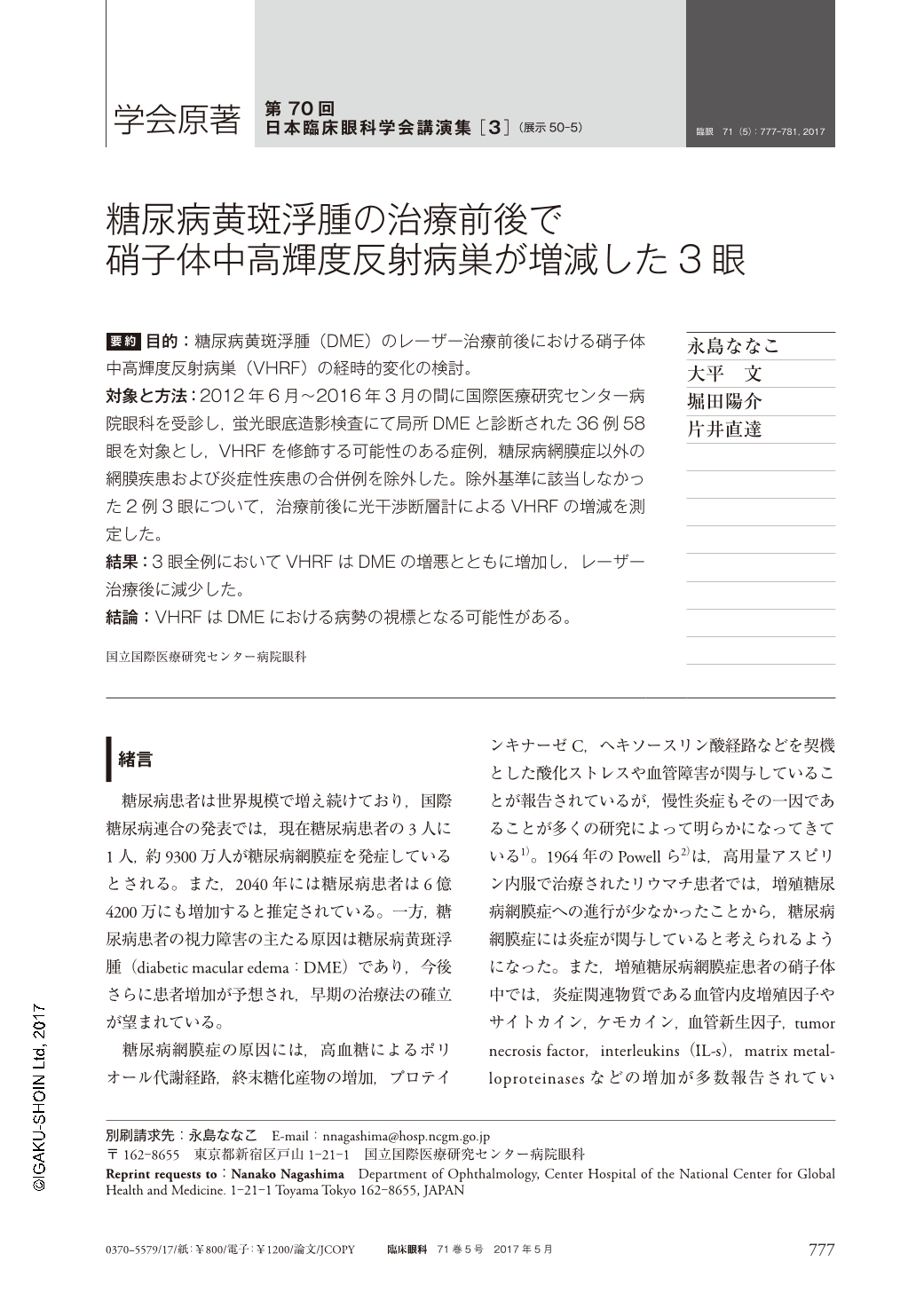
要約 目的:糖尿病黄斑浮腫(DME)のレーザー治療前後における硝子体中高輝度反射病巣(VHRF)の経時的変化の検討。
対象と方法:2012年6月〜2016年3月の間に国際医療研究センター病院眼科を受診し,蛍光眼底造影検査にて局所DMEと診断された36例58眼を対象とし,VHRFを修飾する可能性のある症例,糖尿病網膜症以外の網膜疾患および炎症性疾患の合併例を除外した。除外基準に該当しなかった2例3眼について,治療前後に光干渉断層計によるVHRFの増減を測定した。
結果:3眼全例においてVHRFはDMEの増悪とともに増加し,レーザー治療後に減少した。
結論:VHRFはDMEにおける病勢の視標となる可能性がある。
Abstract Purpose: This study is to report the change in number of hyperreflective foci in eyes with diabetic macula edema before and after laser treatment for microaneurysms.
Cases and Method: Subjects of 58 eyes 36 patients diagnosed as local diabetic macula edema by fluoresceine angiography between June 2012 and March 2016 were evaluated for hyperreflective foci on optical coherence tomography. Eyes with conditions that may affect numbers of hyperreflective foci were excluded. Eyes which required more than 10 shots of laser treatment were also excluded to take into account the possible increase in inflammation due to laser treatment itself. Three eyes of two subjects fulfilled these criteria and were observed for change in number of hyperreflective foci before and after laser treatment.
Results: All 3 eyes showed increase in number of hyperreflective foci with diabetic macula edema exacerbation, and its decrease after laser treatment with resolution of diabetic macula edema.
Conclusion: Number of hyperreflective foci corresponded to the degree of diabetic macula edema before and after laser treatment. hyperreflective foci may become a reliable index for evaluating the severity of diabetic macula edema.

Copyright © 2017, Igaku-Shoin Ltd. All rights reserved.


