Japanese
English
- 有料閲覧
- Abstract 文献概要
- 1ページ目 Look Inside
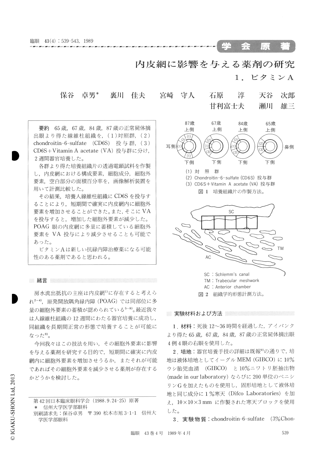
65歳,67歳,84歳,87歳の正常屍体摘出眼より得た線維柱組織を,(1)対照群,(2)chondroitin-6-sulfate (CD6S)投与群,(3)CD6S+Vitamin A acetate (VA)投与群に分け,2週間器官培養した。
各群より得た培養組織片の透過電顕試料を作製し,内皮網における構成要素,細胞成分,細胞外要素,空白部分の面積百分率を,画像解析装置を用いて計測比較した。
その結果,培養人線維柱組織にCD6Sを投与することにより,短期間で確実に内皮網内に細胞外要素を増加させることができた。また,そこにVAを投与すると,増加した細胞外要素が減少した。POAG眼の内皮網に多量に蓄積している細胞外要素をVA投与により減少させることも可能であった。
ビタミンAは新しい抗緑内障治療薬になる可能性のある薬剤であると思われる。
We cultured the trabecular meshwork for 2 weeks using solid agar method. The tissue was obtained from 4 eyes postmortem aged 65, 67, 84 and 87 years each. The cultured specimens were divided into 3 groups: 1) control group, 2) chon-droitin-6-sulfate (CD6S) added group, 3) CD6S + Vitamin A acetate (VA) added group.
The samples were then prepared for transmission electron microscopy. We measured the percentage of areas of cellular space, extracellular material space and empty space in the endothelial meshworkusing computer-assisted morphometry.
When examined 2 weeks after addition of CD6S, all the specimens showed significant increases in extracellular material followed by decrease of empty space in the endothelial meshwork. A signi-ficant decrease of extracellular material and an increase in empty space occurred one day after addition of VA.
A similar decrease of extracellular material was seen in cultured specimen obtained by trabeculectomy after addition of VA.
The finding seemed to suggest that Vitamin A may directly influence the trabecular meshwork cells with a potential of clinical application.

Copyright © 1989, Igaku-Shoin Ltd. All rights reserved.


