Japanese
English
- 有料閲覧
- Abstract 文献概要
- 1ページ目 Look Inside
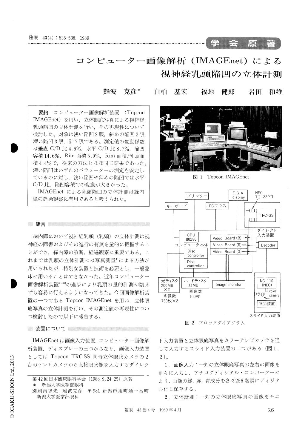
コンピューター画像解析装置(Topcon IMAGEnet)を用い,立体眼底写真による視神経乳頭陥凹の立体計測を行い,その再現性について検討した。対象は浅い陥凹2眼,斜めの陥凹2眼,深い陥凹3眼,計7眼である。測定値の変動係数は垂直C/D比4.6%,水平C/D比8.7%,陥凹容積14.6%,Rim面積5.0%,Rim面積/乳頭面積4.4%で,従来の方法とほぼ同じ結果であった。深い陥凹はいずれのパラメーターの測定も安定しているのに対し,浅い陥凹や斜めの陥凹では水平C/D比,陥凹容積での変動が大きかった。
IMAGEnetによる乳頭陥凹の立体計測は緑内障の経過観察に有用であると考えられた。
We evaluated the reproducibility of optic disc cup measurements as processed by a computerized image analyzer, IMAGEnet (Topcon). We used simulaneous stereoscopic fundus photographs from 7 eyes. Two eyes manifested shallow cup, 2 tempo-ral cup and 3 deep cup. We took ten stereo-photographs for each eye at the same session. Each stereophotograph underwent ten repeated measure-ments.
We found the following values as mean coeffi-cients of variation ± standard deviation: 4.6±2.7%for vertical C/D,4.4±1.8%for rim area/disc area,5.0±1.6%for rim area,8.7±5.0%for horizontal C/D,and 14.6±6.8%for cup volume.Coefficients of vari-ation for horizontal C/D and cup volume weregreater in eyes with shallow or temporal cup thanin those with deep cup.
These results show that measurements of optic disc cupping with the present system can be reason-ably useful in the follow-up of disc changes in gaulcoma.

Copyright © 1989, Igaku-Shoin Ltd. All rights reserved.


