Japanese
English
- 有料閲覧
- Abstract 文献概要
- 1ページ目 Look Inside
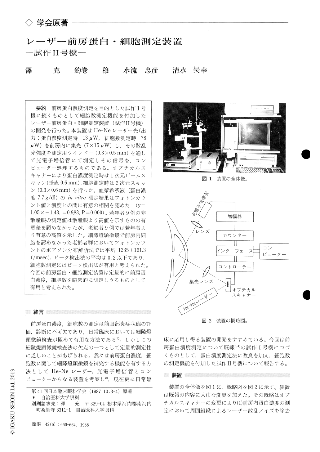
前房蛋白濃度測定を目的とした試作I号機に続くものとして細胞数測定機能を付加したレーザー前房蛋白・細胞測定装置(試作II号機)の開発を行った.本装置はHe-Neレーザー光(出力:蛋白濃度測定時13μW,細胞数測定時78μW)を前房内に集光(7×15μW)し,その散乱光強度を測定用ウインドー(0.3×0.5mm)を通して光電子増倍管にて測定しその信号を,コンピューター処理するものである.オプチカルスキャナーにより蛋白濃度測定時は1次元ビームスキャン(垂直0.6mm),細胞測定時は2次元スキャン(0.3×0.6mm)を行った.血漿希釈液(蛋白濃度7.7g/dl)のin vitro測定結果はフォトンカウント値と濃度との間に有意の相関を認めた(y=1.05×−1.43,=0.983,P=0.000).若年者9例の非散瞳眼の測定値は散瞳眼より高値を示すものの有意差を認めなかったが,老齢者9例では若年者より有意の高値を示した.細隙燈顕微鏡で前房内細胞を認めなかった老齢者群においてフォトンカウントのボアソン分布解析法では平均1235±161.3(/msec),ピーク検出法の平均は0.2以下であり,細胞数測定にはピーク検出法が有用と考えられた.今回の前房蛋白・細胞測定装置は定量的に前房蛋白濃度,細胞数を臨床的に測定しうるものとして有用と考えられた.
We developed an instrument for in vivo quantita-tion of protein concentration and cells in the ante-rior chamber. It consists of three main compo-nents : illumination, measuring device and data processing system.
For the illumination, an He-Ne laser beam is used, with the size of 7×10μm at the place of focus. A photon-counting photomultiplier measures the intensity of scattered beam in the aqueous through a sampling window 0.3×0.5 mm in size. A personal computer controls the system and ana-lyzes the detected signals.
The system has dual modes for measurement of protein concentration and the number of cells. In protein concentration mode, the laser beam is set at 13μW in power output to scan vertically for 0.6 mm. It takes 1 second for the measurement. In cell -count mode, the laser beam is set at 78μW to scan two-dimensionally, 0.6×0.3 mm.It takes 1.2 secondsfor the measurement.
We found a good correlation between the thus measured value and the actual one using human plasma (r=0.993, p=0.00). We also confirmed a good correlation regarding the cell counting mode between the measured value and slit-lamp micro-scopic findings in senile subjects.
In normal eyes in young subjects, values for protein concentration measured 0.13ア0.056 (meanアSD/msec) under mydriatics and 0.15+0. 046 without, corresponding to 3.33 and 3.85 mg/dl. The difference was not significant. Eyes in senile subjects showed significantly higher values, 0.33+0. 15 (p<0.05).
The present system promises to be of clinical value in quantitative measurement of protein con-centration and cells in the anterior chamber when further refined.
Rinsho Ganka (Jpn J Clin Ophthalmol) 42(6) : 660-664, 1988

Copyright © 1988, Igaku-Shoin Ltd. All rights reserved.


