Japanese
English
- 有料閲覧
- Abstract 文献概要
- 1ページ目 Look Inside
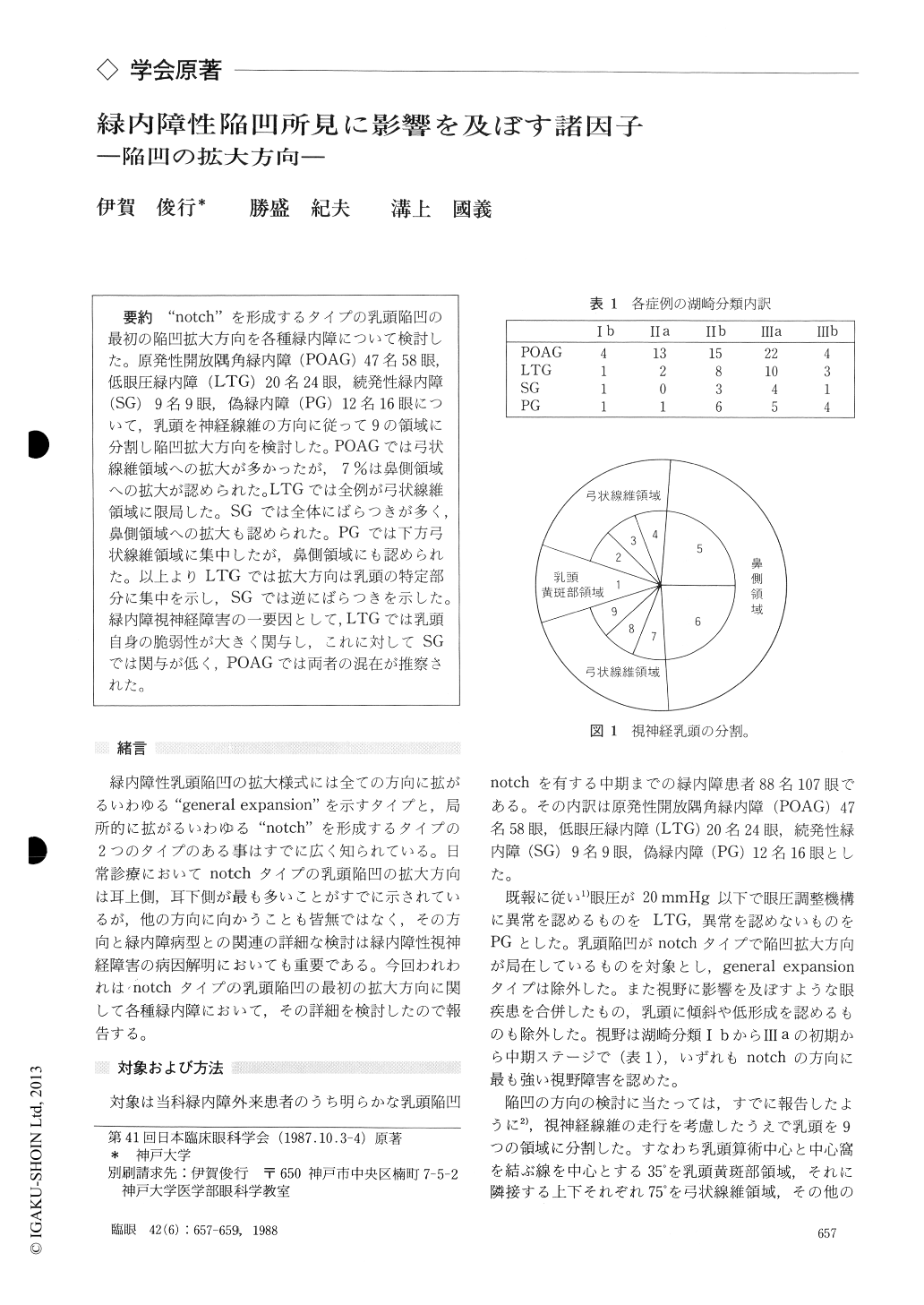
"notch"を形成するタイプの乳頭陥凹の最初の陥凹拡大方向を各種緑内障について検討した.原発性開放隅角緑内障(POAG)47名58眼,低眼圧緑内障(LTG)20名24眼,続発性緑内障(SG)9名9眼,偽緑内障(PG)12名16眼について,乳頭を神経線維の方向に従って9の領域に分割し陥凹拡大方向を検討した.POAGでは弓状線維領域への拡大が多かったが,7%は鼻側領域への拡大が認められた.LTGでは全例が弓状線維領域に限局した.SGでは全体にばらつきが多く,鼻側領域への拡大も認められた.PGでは下方弓状線維領域に集中したが,鼻側領域にも認められた.以上よりLTGでは拡大方向は乳頭の特定部分に集中を示し,SGでは逆にばらつきを示した.緑内障視神経障害の一要因として,LTGでは乳頭自身の脆弱性が大きく関与し,これに対してSGでは関与が低く,POAGでは両者の混在が推察された.
We evaluated the optic disc in 107 eyes with glaucoma or allied diseases : primary open angle glaucoma (POAG) 58 eyes, low tension glaucoma (LTG) 24, secondary glaucoma (SG) 9, and pseudoglaucoma (PG) 16.
We divided the optic disc into 9 areas in accor-dance to the distributional pattern of retinal nerve fiber bundles.
In all the cases of LTG, the most significant enlargement of cupping was located in the upperand lower temporal disc areas. In 81% of eyes with PG, the cupping enlarged in the disc area corre-sponding to the arcuate nerve fiber. Enlargement of cupping showed no definite pattern in eyes with SG.The enlargement developed in the arcuate nerve fiber disc area in 93% of eyes with POAG, and toward the nasal disc area in the other 7%. The findings seemed to imply the vulnerability of the optic disc in LTG. The supposed vulnerability seemed to be of lesser order in POAG and to a negligible degree in SG.
Rinsho Ganka (Jpn J Clin Ophthalmol) 42(6) : 657-659, 1988
Copyright © 1988, Igaku-Shoin Ltd. All rights reserved.