Japanese
English
- 有料閲覧
- Abstract 文献概要
- 1ページ目 Look Inside
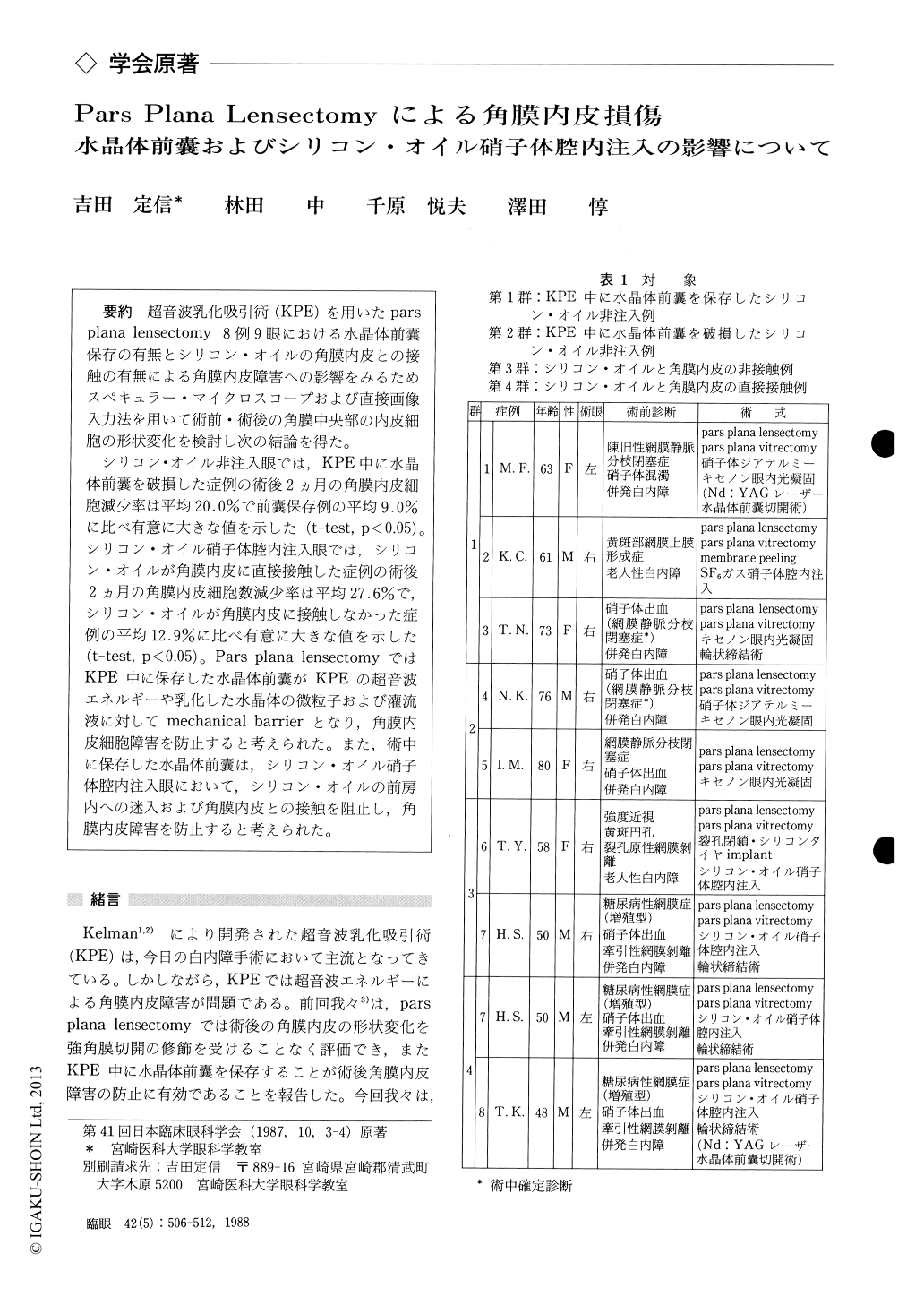
超音波乳化吸引術(KPE)を用いたparsplana lensectomy 8例9眼における水晶体前嚢保存の有無とシリコン・オイルの角膜内皮との接触の有無による角膜内皮障害への影響をみるためスペキュラー・マイクロスコープおよび直接画像入力法を用いて術前・術後の角膜中央部の内皮細胞の形状変化を検討し次の結論を得た.
シリコン・オイル非注入眼では,KPE中に水晶体前嚢を破損した症例の術後2カ月の角膜内皮細胞減少率は平均20.0%で前嚢保存例の平均9.0%に比べ有意に大きな値を示した(t-test, p<0.05).シリコン・オイル硝子体腔内注入眼では,シリコン・オイルが角膜内皮に直接接触した症例の術後2カ月の角膜内皮細胞数減少率は平均27.6%で,シリコン・オイルが角膜内皮に接触しなかつた症例の平均12.9%に比べ有意に大きな値を示した(t-test, p<0.05).Pars plana lensectomyではKPE中に保存した水晶体前嚢がKPEの超音波エネルギーや乳化した水晶体の微粒子および灌流液に対してmechanical barrierとなり,角膜内皮細胞障害を防止すると考えられた.また,術中に保存した水晶体前嚢は,シリコン・オイル硝子体腔内注入眼において,シリコン・オイルの前房内への迷入および角膜内皮との接触を阻止し,角膜内皮障害を防止すると考えられた.
We evaluated the damage to the corneal endoth-elium after pars plana lensectomy by phacoemul-sification in 9 eyes, 8 patients. We analyzed the endothelium in the central cornea before and after surgery, using specular microscope and computer -assisted image analyzer.
In eyes without additional intravitreal liquid silicone, mean corneal endothelial cell loss was 9. 0% when the anterior lens capsule was kept intact during phacoemulsification. The mean cell loss increased to 20.0% when the anterior lens capsulewas ruptured during phacoemulsification.
In eyes with additional intravitreal liquid sili-cone, mean corneal endothelial cell loss was 12.9% when the liquid silicone was not in contact with the corneal endothelium. The mean cell loss increased, significantly, to 27.6% when the liquid silicone was in contact with the corneal endothelium.
These findings suggest that the anterior lens capsule acts as a mechanical barrier, preventing the corneal endothelium from harmful effects by the ultrasonic energy, fragmented lens particles and irrigation fluid during phacoemulsification. An intact anterior lens capsule is of another benefit as it keeps the intravitreal liquid silicone away from the anterior chamber and the corneal endothelium.
Rinsho Ganka (Jpn J Clin Ophthalmol) 42(5) : 506-512, 1988

Copyright © 1988, Igaku-Shoin Ltd. All rights reserved.


