Japanese
English
- 有料閲覧
- Abstract 文献概要
- 1ページ目 Look Inside
緒言
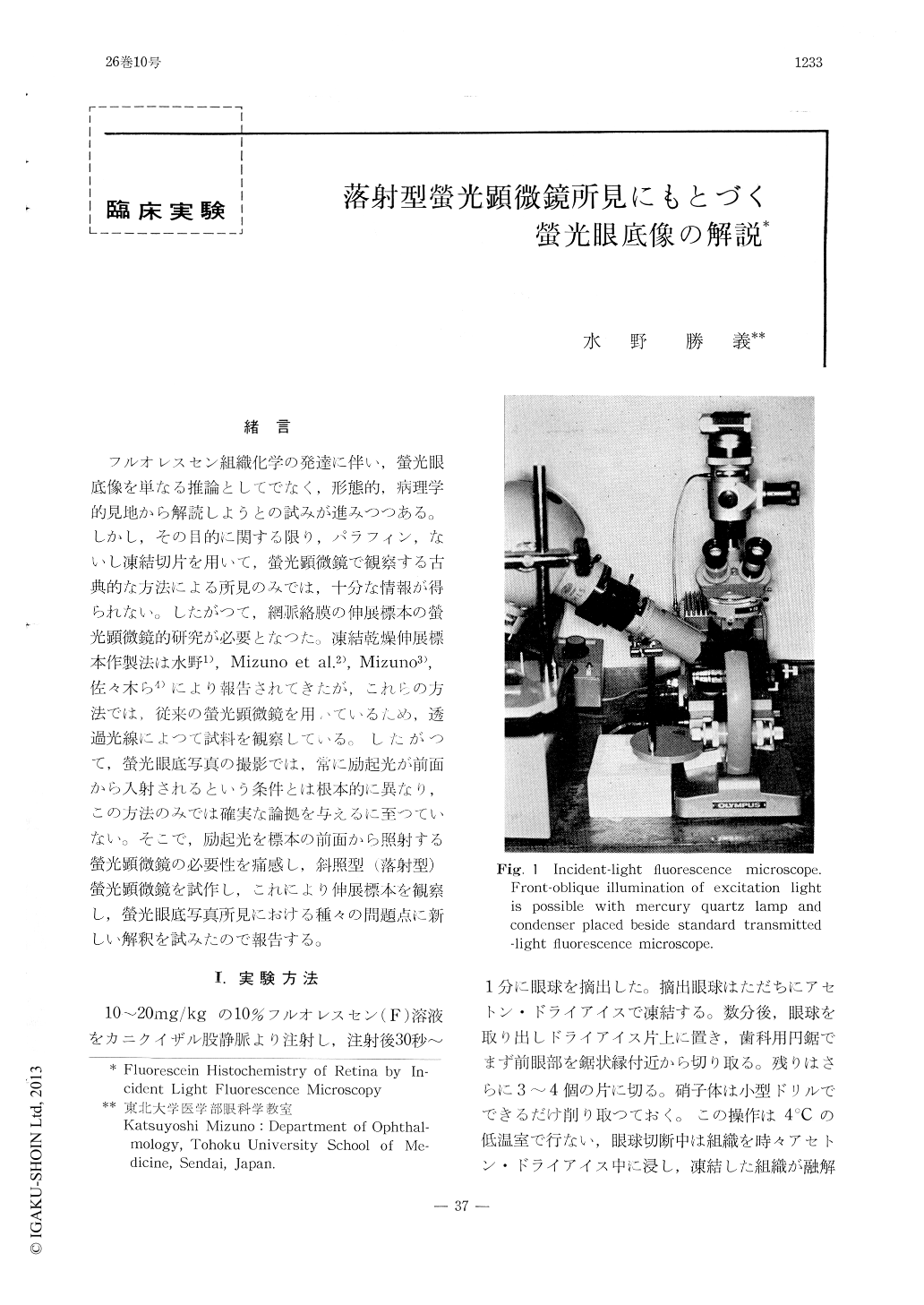
フルオレスセン組織化学の発達に伴い,螢光眼底像を単なる推論としてでなく,形態的,病理学的見地から解読しようとの試みが進みつつある。しかし,その目的に関する限り,パラフィン,ないし凍結切片を用いて,螢光顕微鏡で観察する古典的な方法による所見のみでは,十分な情報が得られない。したがつて,網脈絡膜の伸展標本の螢光顕微鏡的研究が必要となつた。凍結乾燥伸展標本作製法は水野1),Mizuno et al.2),Mizuno3),佐々木ら4)により報告されてきたが,これらの方法では,従来の螢光顕微鏡を用いているため,透過光線によつて試料を観察している。したがつて,螢光眼底写真の撮影では,常に励起光が前面から入射されるという条件とは根本的に異なり,この方法のみでは確実な論拠を与えるに至つていない。そこで,励起光を標本の前面から照射する螢光顕微鏡の必要性を痛感し,斜照型(落射型)螢光顕微鏡を試作し,これにより伸展標本を観察し,螢光眼底写真所見における種々の問題点に新しい解釈を試みたので報告する。
The localization of fluorescein in the whole mounted retina and choroid of rhesus monkey was studied by using the incident-light fluore-scence microscope. The findings were compared with those by routine fluorescein angiography in humans.
The macular avascular area lacked fluores-cence regardless of the presence or absence of the pigment epithelium and/or the choroid. Fur-ther, no significant differences were found in the intensity of background fluorescence in retinal preparations with or without the pigment epi-thelium.

Copyright © 1972, Igaku-Shoin Ltd. All rights reserved.


