Japanese
English
- 有料閲覧
- Abstract 文献概要
- 1ページ目 Look Inside
緒言
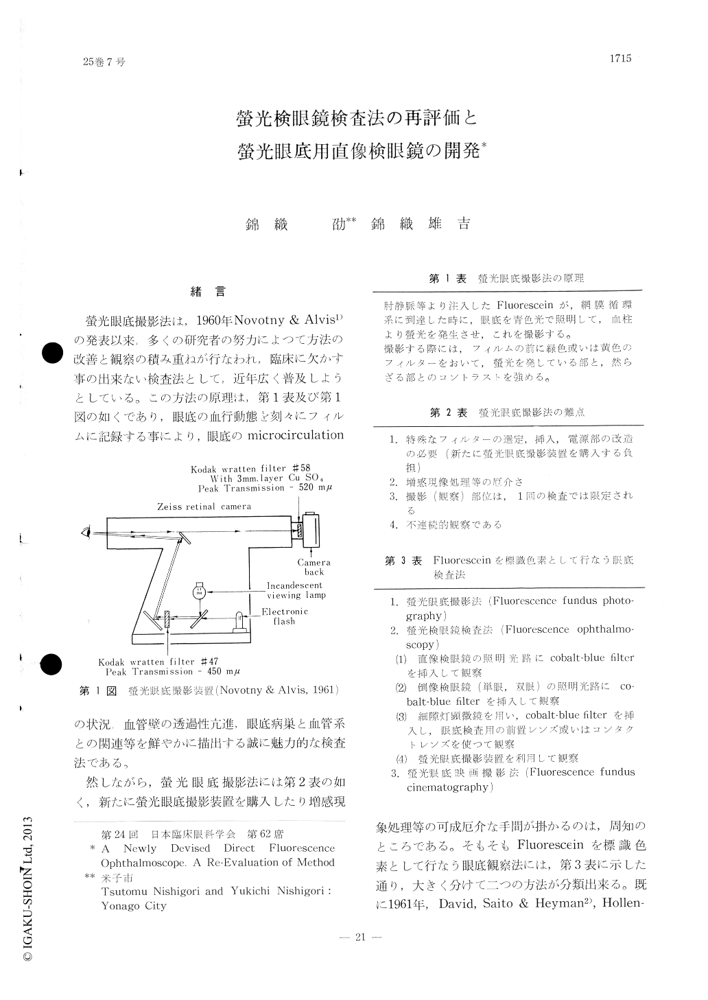
螢光眼底撮影法は,1960年Novotny & Alvis1)の発表以来,多くの研究者の努力によつて方法の改善と観察の積み重ねが行なわれ,臨床に欠かす事の出来ない検査法として,近年広く普及しようとしている。この方法の原理は,第1表及び第1図の如くであり,眼底の血行動態を刻々にフィルムに記録する事により,眼底のmicrocirculationの状況,血管壁の透過性亢進,眼底病巣と血管系との関連等を鮮やかに描出する誠に魅力的な検査法である。
然しながら,螢光眼底撮影法には第2表の如く,新たに螢光眼底撮影装置を購入したり増感現象処理等の可成厄介な手間が掛かるのは,周知のところである。そもそもFluoresceinを標識色素として行なう眼底観察法には,第3表に示した通り,大きく分けて二つの方法が分類出来る。既に1961年,David, Saito & Heyman2), Hollen—horst & Kearns3),1964年にはPemberton &Britton4)が,肘静脈—網膜循環時間の測定に,co—balt-blue filterを入れた直像鏡を用いている。
A new direct fluorescence ophthalmoscope has been devised by the authors by inserting a co-balt-blue glass filter (Hoya B-390) in the illu-minating system and a blue-absorbing yellow filter (Hoya Y-50) in the observing system of the direct ophthalmoscope. As the light source, the hitherto current incandescent lamp proved to be sufficient so that there was no necessity to use glass fiber system.
Fifty-one cases, including central serous re-tinopathy, diabetic retinopathy and obstruction of the retinal vein, have been examined with the direct fluorescence ophthalmoscope.

Copyright © 1971, Igaku-Shoin Ltd. All rights reserved.


