Japanese
English
特集 第22回日本臨床眼科学会講演集 (その4)
螢光眼底撮影法による糖尿病性網膜症の観察—(第2報)
An Investigation on the Fluorescein Fundus Angiogrophy of the Diabetic Retinopathy
茂木 劼
1
,
山上 磐
1
,
葛谷 信貞
2
,
尾形 安三
2
,
平塚 任
2
,
儀間 祺昇
2
Tsutomu Motegi
1
,
Iwao Yamagami
1
,
Nobusada Kuzuya
2
,
Yasuzo Ogata
2
,
Atsushi Hiratsuka
2
,
Yoshinori Gima
2
1虎の門病院眼科
2虎の門病院内分泌科
1Department of Ophthalmology, Toranomon Hospital
2Department of Endocrinology, Toranomon Hospital
pp.523-528
発行日 1969年4月15日
Published Date 1969/4/15
DOI https://doi.org/10.11477/mf.1410204061
- 有料閲覧
- Abstract 文献概要
- 1ページ目 Look Inside
I.緒言
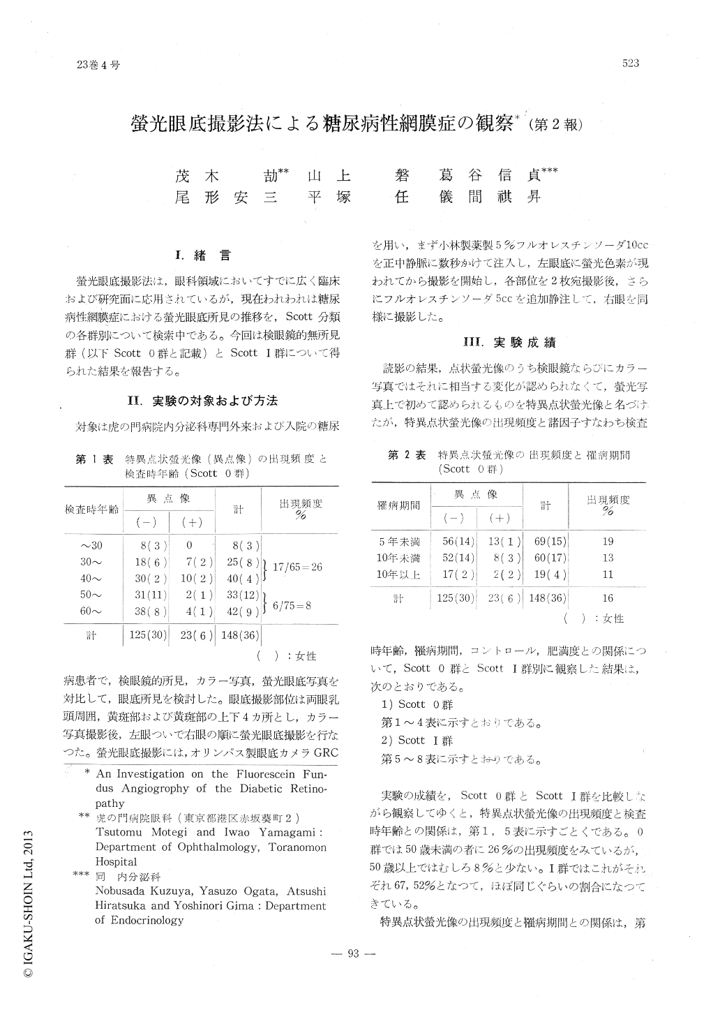
螢光眼底撮影法は,眼科領域においてすでに広く臨床および研究面に応用されているが,現在われわれは糖尿病性網膜症における螢光眼底所見の推移を,Scott分類の各群別について検索中である。今回は検眼鏡的無所見群(以下Scott 0群と記載)とScottⅠ群について得られた結果を報告する。
Fluorescein fundus angiography was conduct-ed on 185 patients with diabetic retinopathy belonging to Stage 0 and I according to Scott. As a conspicuous recurrent occurrence, isolated fluorescent dots were detected in fundus areas which seemed normal upon ordinary color pho-tographs. This phenomenon was detected in 54% of 37 patients belonging to Stage 1 and seemed to be correlated with the severity of the re-tinopathy rather than the duration of the dia-betes.

Copyright © 1969, Igaku-Shoin Ltd. All rights reserved.


