Japanese
English
〔眼・光学学会〕
試作眼球開壁術用手術顕微鏡
An Operating Microscope for Bulbotomy
奥田 観士
1
,
那須 欽爾
1
Kanji Okuda
1
,
Kinji Nasu
1
1岡山大学医学部眼科学教室
1Department of Ophthalmology, Okayama University,Medical School
pp.795-797
発行日 1967年6月15日
Published Date 1967/6/15
DOI https://doi.org/10.11477/mf.1410203680
- 有料閲覧
- Abstract 文献概要
- 1ページ目 Look Inside
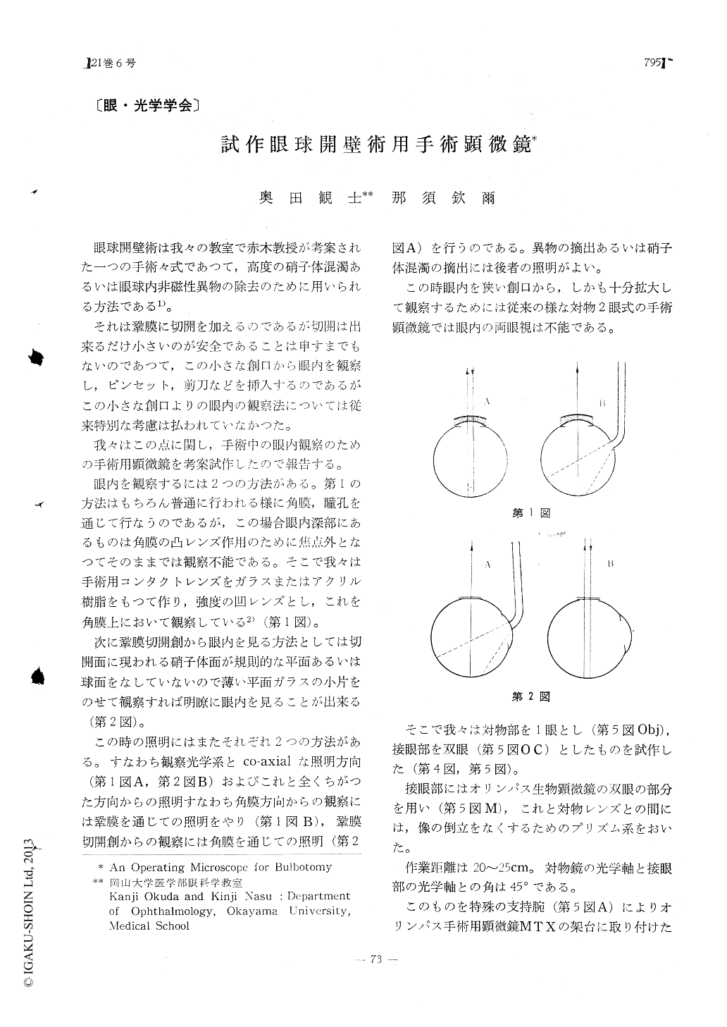
眼球開壁術は我々の教室で赤木教授が考案された一つの手術々式であつて,高度の硝子体混濁あるいは眼球内非磁性異物の除去のために用いられる方法である1)。
それは鞏膜に切開を加えるのであるが切開は出来るだけ小さいのが安全であることは申すまでもないのであつて,この小さな創口から眼内を観察し,ピンセット,剪刀などを挿入するのであるがこの小さな創口よりの眼内の観察法については従来特別な考慮は払われていなかつた。
Bulbotomy is an operation to open the eye-ball to remove dense vitreous opacities or intra-ocular non-magnetic foreign bodies. This mi-croscope enables the operator to examine the interior of the eyeball through a very narrow scleral incision. The microscope has one objec-tive lens and a pair of eye pieces.

Copyright © 1967, Igaku-Shoin Ltd. All rights reserved.


