Japanese
English
- 有料閲覧
- Abstract 文献概要
- 1ページ目 Look Inside
I.緒論
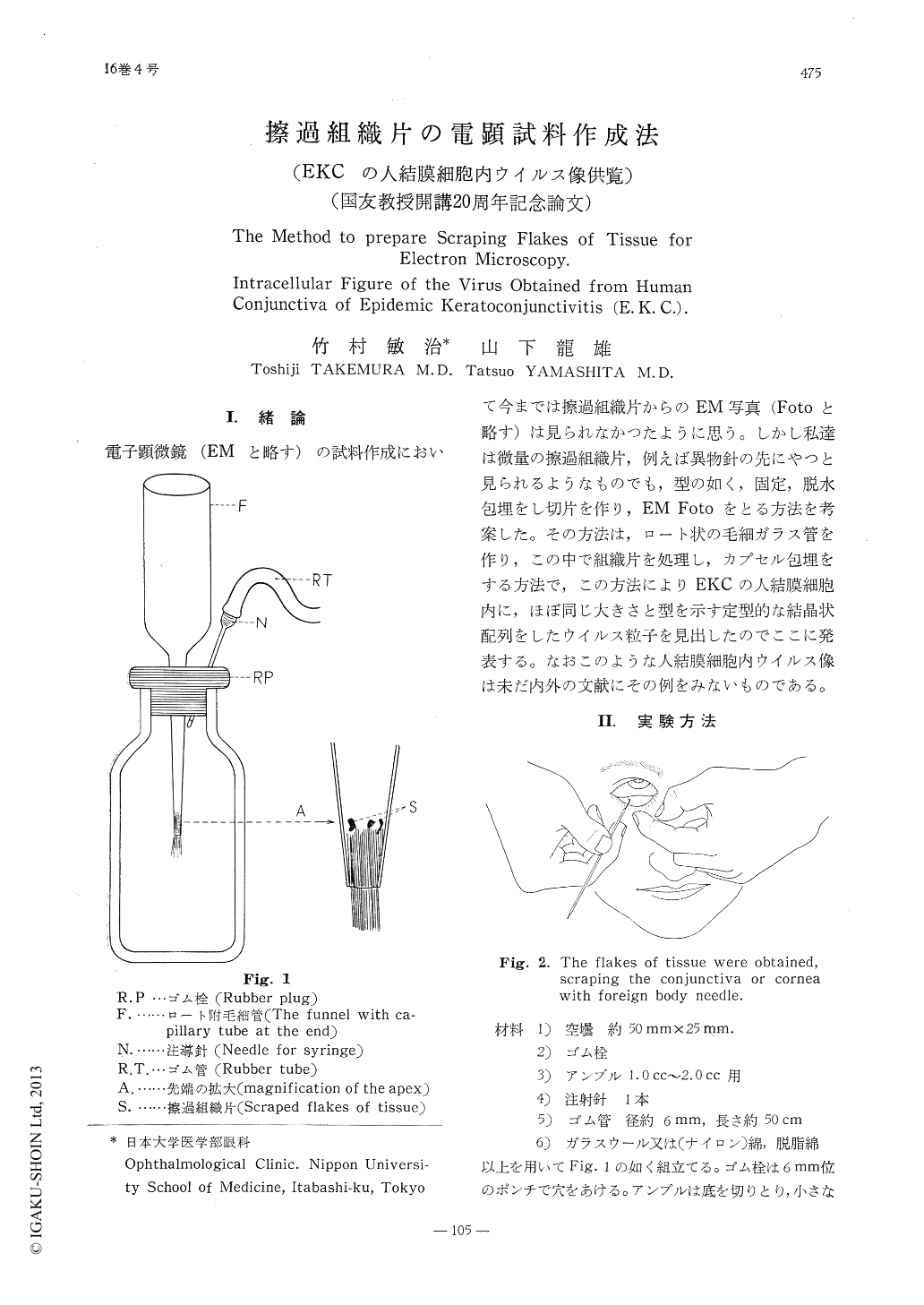
電子顕微鏡(EMと略す)の試料作成において今までは擦過組織片からのEM写真(Fotoと略す)は見られなかつたように思う。しかし私達は微量の擦過組織片,例えば異物針の先にやつと見られるようなものでも,型の如く,固定,脱水包埋をし切片を作り,EM Fotoをとる方法を考案した。その方法は,ロート状の毛細ガラス管を作り,この中で組織片を処理し,カプセル包埋をする方法で,この方法によりEKCの人結膜細胞内に,ほぼ同じ大きさと型を示す定型的な結晶状配列をしたウイルス粒子を見出したのでここに発表する。なおこのような人結膜細胞内ウイルス像は未だ内外の文献にその例をみないものである。
1. Preamble
In regard to the preparation for electron microscopy (EM), various methods were re-ported. However, no description was found in regard to scraping flakes of tissue. In this report, the authors introduced a method to take an electron microscopic photograph (E M Poto), making the preparation from the minutest scraping flakes, through usual route of fixation, dehydration and envelopment. The method is as follows. A glass funnel with capillary tube at the end is prepared.

Copyright © 1962, Igaku-Shoin Ltd. All rights reserved.


