Japanese
English
- 有料閲覧
- Abstract 文献概要
- 1ページ目 Look Inside
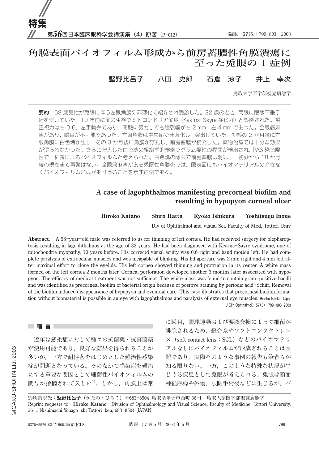
要約 58歳男性が兎眼に伴う左眼角膜の菲薄化で紹介され受診した。32歳のとき,両眼に眼瞼下垂手術を受けていた。10年前に筋の生検でミトコンドリア筋症(Kearns-Sayre症候群)と診断された。矯正視力は右0.6,左手動弁であり,閉瞼に努力しても瞼裂幅が右2mm,左4mmであった。全眼筋麻痺があり,瞬目が不可能であった。左眼角膜は中央部で菲薄化し,突出していた。初診の2か月後に左眼角膜に白色塊が生じ,その3か月後に角膜が穿孔し,前房蓄膿が続発した。薬物治療では十分な効果が得られなかった。さらに増大した白色塊の組織学的検索でグラム陽性の桿菌が検出され,PAS染色陽性で,細菌によるバイオフィルムと考えられた。白色塊の除去で前房蓄膿は消退し,初診から18か月後の現在まで再発はない。全眼筋麻痺がある兎眠性角膜炎では,眼表面にもバイオマテリアルの介在なくバイオフィルム形成がありうることを示す症例である。
Abstract. A 58-year-old male was referred to us for thinning of left cornea. He had received surgery for blepharoptosis resulting in lagophthalmos at the age of 32 years. He had been diagnosed with Kearns-Sayre syndrome,one of mitochondria myopathy,10 years before. His correctd visual acuity was 0.6 right and hand motion left. He had complete paralysis of extraocular muscles and was incapable of blinking. His lid aperture was 2 mm right and 4mm left after maximal effort to close the eyelids. His left cornea showed thinning and protrusion in its center. A white mass formed on the left cornea 2 months later. Corneal perforation developed another 3 months later associated with hypopyon. The efficacy of medical treatment was not sufficient. The white mass was found to contain gram-positive bacilli and was identified as precorneal biofilm of bacterial origin because of positive staining by periodic acid-Schiff. Removal of the biofilm induced disappearance of hypopyon and eventual cure. This case illustrates that precorneal biofilm formation without biomaterial is possible in an eye with lagophthalmos and paralysis of external eye muscles.

Copyright © 2003, Igaku-Shoin Ltd. All rights reserved.


