Japanese
English
- 有料閲覧
- Abstract 文献概要
- 1ページ目 Look Inside
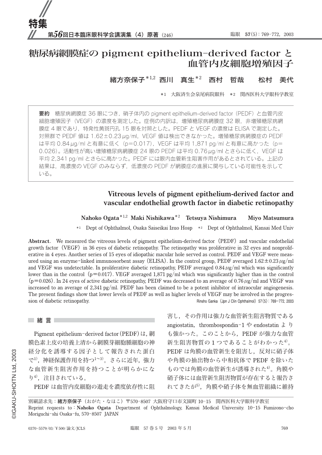
要約 糖尿病網膜症36眼につき,硝子体内のpigment epithelium-derived factor(PEDF)と血管内皮細胞増殖因子(VEGF)の濃度を測定した。症例の内訳は,増殖糖尿病網膜症32眼,非増殖糖尿病網膜症4眼であり,特発性黄斑円孔15眼を対照とした。PEDFとVEGFの濃度はELISAで測定した。対照群でPEDF値は1.62±0.23μg/ml,VEGF値は検出できなかった。増殖糖尿病網膜症のPEDFは平均0.84μg/mlと有意に低く(p=0.017),VEGFは平均1,871pg/mlと有意に高かった(p=0.026)。活動性が高い増殖糖尿病網膜症24眼のPEDFは平均0.76μg/mlとさらに低く,VEGFは平均2,341pg/mlとさらに高かった。PEDFには眼内血管新生阻害作用があるとされている。上記の結果は,高濃度のVEGFのみならず,低濃度のPEDFが網膜症の進展に関与している可能性を示している。
Abstract. We measured the vitreous levels of pigment epithelium-derived factor(PEDF)and vascular endothelial growth factor(VEGF)in 36 eyes of diabetic retinopathy. The retinopathy was proliferative in 32 eyes and nonproliferative in 4 eyes. Another series of 15 eyes of idiopathic macular hole served as control. PEDF and VEGF were measured using an enzyme-linked immunosorbent assay(ELISA). In the control group,PEDF averaged 1.62±0.23μg/ml and VEGF was undetectable. In proliferative diabetic retinopathy,PEDF averaged 0.84μg/ml which was significantly lower than in the control(p=0.017). VEGF averaged 1,871pg/ml which was significantly higher than in the control(p=0.026). In 24 eyes of active diabetic retinopathy,PEDF was decreased to an average of 0.76μg/ml and VEGF was increased to an average of 2,341pg/ml. PEDF has been claimed to be a potent inhibitor of intraocular angiogenesis. The present findings show that lower levels of PEDF as well as higher levels of VEGF may be involved in the progression of diabetic retinopathy.

Copyright © 2003, Igaku-Shoin Ltd. All rights reserved.


