Japanese
English
- 有料閲覧
- Abstract 文献概要
- 1ページ目 Look Inside
- 参考文献 Reference
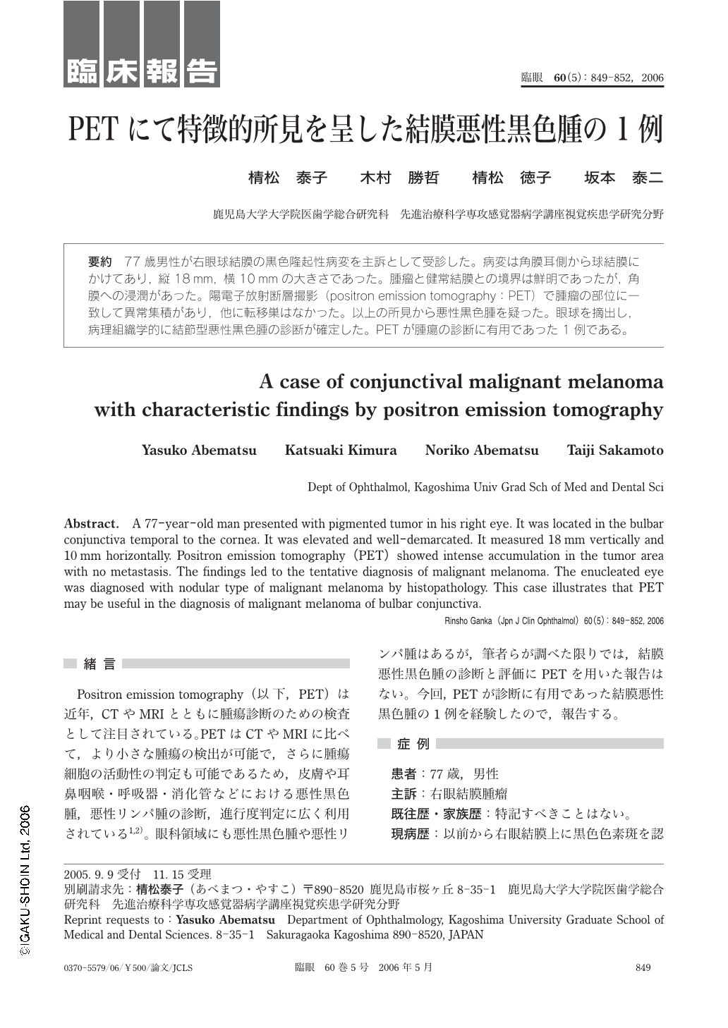
77歳男性が右眼球結膜の黒色隆起性病変を主訴として受診した。病変は角膜耳側から球結膜にかけてあり,縦18mm,横10mmの大きさであった。腫瘤と健常結膜との境界は鮮明であったが,角膜への浸潤があった。陽電子放射断層撮影(positron emission tomography:PET)で腫瘤の部位に一致して異常集積があり,他に転移巣はなかった。以上の所見から悪性黒色腫を疑った。眼球を摘出し,病理組織学的に結節型悪性黒色腫の診断が確定した。PETが腫瘍の診断に有用であった1例である。
A 77-year-old man presented with pigmented tumor in his right eye. It was located in the bulbar conjunctiva temporal to the cornea. It was elevated and well-demarcated. It measured 18 mm vertically and 10 mm horizontally. Positron emission tomography(PET)showed intense accumulation in the tumor area with no metastasis. The findings led to the tentative diagnosis of malignant melanoma. The enucleated eye was diagnosed with nodular type of malignant melanoma by histopathology. This case illustrates that PET may be useful in the diagnosis of malignant melanoma of bulbar conjunctiva.

Copyright © 2006, Igaku-Shoin Ltd. All rights reserved.


