Japanese
English
- 有料閲覧
- Abstract 文献概要
- 1ページ目 Look Inside
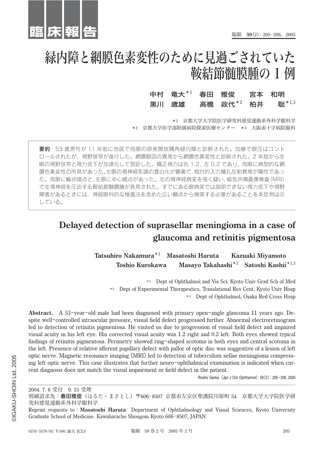
53歳男性が11年前に他医で両眼の原発開放隅角緑内障と診断された。加療で眼圧はコントロールされたが,視野狭窄が進行した。網膜眼図の異常から網膜色素変性と診断された。2年前から左眼の視野狭窄と視力低下が加速化して受診した。矯正視力は右1.2,左0.2であり,両眼に典型的な網膜色素変性の所見があった。左眼の視神経乳頭の蒼白化が顕著で,相対的入力瞳孔反射異常が陽性であった。両眼に輪状暗点と,左眼に中心暗点があった。左の視神経病変を強く疑い,磁気共鳴画像検査(MRI)で左視神経を圧迫する鞍結節髄膜腫が発見された。すでにある眼病変では説明できない視力低下や視野障害があるときには,神経眼科的な検査法を含めた広い観点から検索する必要があることを本症例は示している。
A 53-year-old male had been diagnosed with primary open-angle glaucoma 11 years ago. Despite well-controlled intraocular pressure,visual field defect progressed further. Abnormal electroretinogram led to detection of retinitis pigmentosa. He visited us due to progression of visual field defect and impaired visual acuity in his left eye. His corrected visual acuity was 1.2 right and 0.2 left. Both eyes showed typical findings of retinitis pigmentosa. Perimetry showed ring-shaped scotoma in both eyes and central scotoma in the left. Presence of relative afferent pupillary defect with pallor of optic disc was suggestive of a lesion of left optic nerve. Magnetic resonance imaging(MRI)led to detection of tuberculum sellae meningioma compressing left optic nerve. This case illustrates that further neuro-ophthalmical examination is indicated when current diagnosis does not match the visual impairment or field defect in the patient.

Copyright © 2005, Igaku-Shoin Ltd. All rights reserved.


