Japanese
English
- 有料閲覧
PPV購入案内
1,320円 (1,200円+税10%) こちらは、1341ページから1346ページの文献です。購入には医書.jp本会員登録が必要です。
会員登録完了後に改めて上記「購入サイトへ」を選択してください。
または「購入サイトへ」選択後に「購入ログイン」、「新規会員登録」の順に進んでください。
会員登録について詳しくはをご確認ください。
- Abstract 文献概要
- 1ページ目 Look Inside
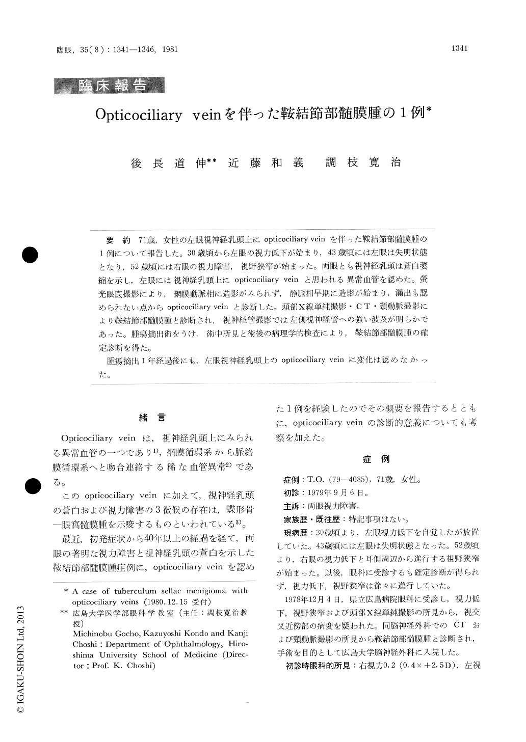
71歳,女性の左眼視神経乳頭上にopticociliary veinを伴った鞍結節部髄膜腫の1例について報告した。30歳頃から左眼の視力低下が始まり,43歳頃には左眼は失明状態となり,52歳頃には右眼の視力障害,視野狭窄が始まった。両眼とも視神経乳頭は蒼白萎縮を示し,左眼には視神経乳頭上にopticociliary veinと思われる異常血管を認めた。螢光眼底撮影により,網膜動脈相に造影がみられず,静脈相早期に造影が始まり,漏出も認められない点からopticociliary veinと診断した。頭部X線単純撮影・CT・頸動脈撮影により鞍結節部髄膜腫と診断され,視神経管撮影では左側視神経管への強い波及が明らかであった。腫瘍摘出術をうけ,術中所見と術後の病理学的検査により,鞍結節部髄膜腫の確定診断を得た。
腫瘍摘出1年経過後にも,左眼視神経乳頭上のopticociliary veinに変化は認めなかった。
A 71-year-old female had a forty-year history of progressive loss of vision in her left eye. Impairment of visual acuity and visual field in her right eye started to progress at the age of 52 years. The visual acuity was now 0.4 RE and 0LE. The optic disc was pale in both eyes. In the left eye, the disc margin was poorly demarcated and the disc vessels were tortuous, which proved to be opticociliary veins by fluo rescein angiography.
A left parasellar tumor was detected by computed tomography. It proved to be transitional menin-gioma on histopathological studies.
Copyright © 1981, Igaku-Shoin Ltd. All rights reserved.