Japanese
English
- 有料閲覧
- Abstract 文献概要
- 1ページ目 Look Inside
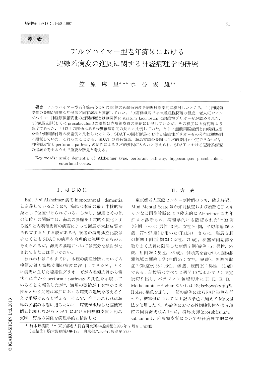
アルツハイマー型老年痴呆(SDAT)33例の辺縁系病変を病理形態学的に検討したところ,1)内嗅領皮質の萎縮が高度な症例ほど固有海馬も萎縮していた,2)固有海馬では神経細胞脱落の程度,老人斑やアルツハイマー神経原線維変化の出現頻度とは無関係にstratum lacunosumに線維性グリオーゼが認められた,3)海馬支脚(とくにprosubiculum)の萎縮は内嗅領皮質の萎縮に比例していたが,その程度は固有海馬より高度であった,4)以上の関係はある程度罹病期間の長さに比例していた。さらに無酸素脳症例と内嗅領皮質を含む側副溝付近の梗塞例と比較したところ,SDATの固有海馬における線維性グリオーゼの分布は梗塞例に類似していた。これらのことから,SDATの固有海馬,海馬支脚の萎縮は1次的要因も否定できないが,内嗅領皮質とperforant pathwayの変性による2次的要因が大きいと考えられ,SDATにおける辺縁系病変の進展を考えるうえで重要な所見と考える。
Neuropathological study on the limbic lesion of 33 autopsy cases with senile dementia of Alzheimer type (SDAT) showed as follows. 1) The entorhinal cortex was more atrophied than the hippocampus. 2) Neuronal loss was found in the 2nd and 3rd layers of the entorhinal cortex, irrespective of the different numbers of senile plaques and neurofibrillary tan-gles (NFTs). 3) Fibrillary gliosis occurred in the stratum lacunosum of the hippocampus, irrespective of the different degrees of neuronal loss in the stratum pyramidale of the hippocampus. 4) The prosubiculum showed gliosis disproportional to neuronal loss.

Copyright © 1997, Igaku-Shoin Ltd. All rights reserved.


