Japanese
English
- 有料閲覧
- Abstract 文献概要
- 1ページ目 Look Inside
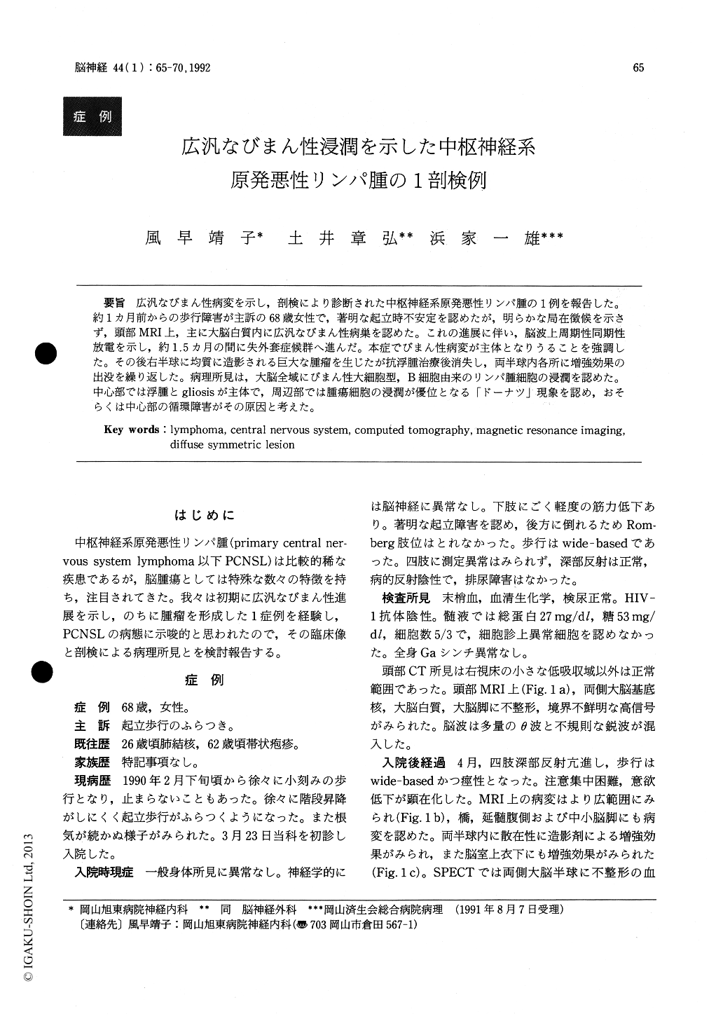
広汎なびまん性病変を示し,剖検により診断された中枢神経系原発悪性リンパ腫の1例を報告した。約1カ月前からの歩行障害が主訴の68歳女性で,著明な起立時不安定を認めたが,明らかな局在徴候を示さず,頭部MRI上,主に大脳白質内に広汎なびまん性病巣を認めた。これの進展に伴い,脳波上周期性同期性放電を示し,約1.5カ月の間に失外套症候群へ進んだ。本症でびまん性病変が主体となりうることを強調した。その後右半球に均質に造影される巨大な腫瘤を生じたが抗浮腫治療後消失し,両半球内各所に増強効果の出没を繰り返した。病理所見は,大脳全域にびまん性大細胞型,B細胞由来のリンパ腫細胞の浸潤を認めた。中心部では浮腫とgliosisが主体で,周辺部では腫瘍細胞の浸潤が優位となる「ドーナツ」現象を認め,おそらくは中心部の循環障害がその原因と考えた。
A case of primary central nervous system lymphoma (PCNSL) who initially showed clinical pictures like encephalitis and diffuse lesions on MRI was reported, including postmortem pathologic examinations. A 68-year-old woman was seen in March, 1990 with a 1-month history of the progresive gait disturbance. She was very unstable and could barely stand by herself, though she did not show any focal neurologic deficits. She showed no evidence of systemic diseases. The cerebrospinal fluid analysis was normal. T2-weighted image of MRI demonstrated the diffuse symmetric hyperintense lesions mainly in the periventricular white matter.
The progressive intellectual decline and the spasticity of four limbs developed as the diffuse lesions on MRI gradually extended. Despite the administration of cor-ticosteroids under the presumptive diagnosis of PCNSL, she rapidly fell into the apallic syndrome within two months. Her EEG showed periodic synchronous dis-charges.
Three months later, she suddenly developed signs of right uncal herniation. CT showed a large mass lesion in the right hemisphere. After the anti-edema therapy, signs of herniation regressed. The serial CT scans demonstrated a gradual decrease in the mass effect, while another multi-nodular lesions appeared and then disappeared one after another bilaterally. Eventually, the diffuse low densities in the cerebral white matter and the ventricular enlargement had remained. She died of bronchopneumonia eight month after the onset of symp-toms. The clinical importance of the diagnosis of PCNSL which initially shows diffuse symmetric lesions without a mass is stressed.
Postmortem examinations revealed PCNSL, diffuse, large cell type according to the Lymphoma Study Group classification. The lymphoma cells were proved to be B cell origin from the immunohistochemical study of fro-zen tissue.
Macroscopically, there was a soft infiltrative lesion in the right centrum simiovale, which extended to subin-sular and frontal white matter, thalamus and lenticular nucleus. Similar lesions were present in the left thalamus, lenticular nucleus and the surrounding white matter. They also involved the hypothalamus and the midbrain.
Microscopic infiltrations of lymphoma cells were far extensive than expected macroscopically. Dense peri-vascular and parenchymatous lymphoma cells were found in the right lenticular nucleus. In the right centrum semiovale, necrotic edema and proliferations of capillaries were surrounded by severe gliosis with sparse lymphoma cells. There were marked infiltrations of lymphoma cells around the focus of gliosis. Similarly in the left basal ganglia, focal areas of necrosis and diffuse gliosis were surrounded by dense lymphoma cells. These distributions of lymphoma cells were like a doughnut, which was the finding commonly seen also in the deep white matter of the both hemispheres. When the lymphoma cells infiltrated along capillaries, a cen-tral ischemia of the tumor, and subsequently, edemas and reactive gliosis might follow. It is conceivable that the ischemia might be related to the marked changes of the CT findings.

Copyright © 1992, Igaku-Shoin Ltd. All rights reserved.


