Japanese
English
- 有料閲覧
- Abstract 文献概要
- 1ページ目 Look Inside
- 参考文献 Reference
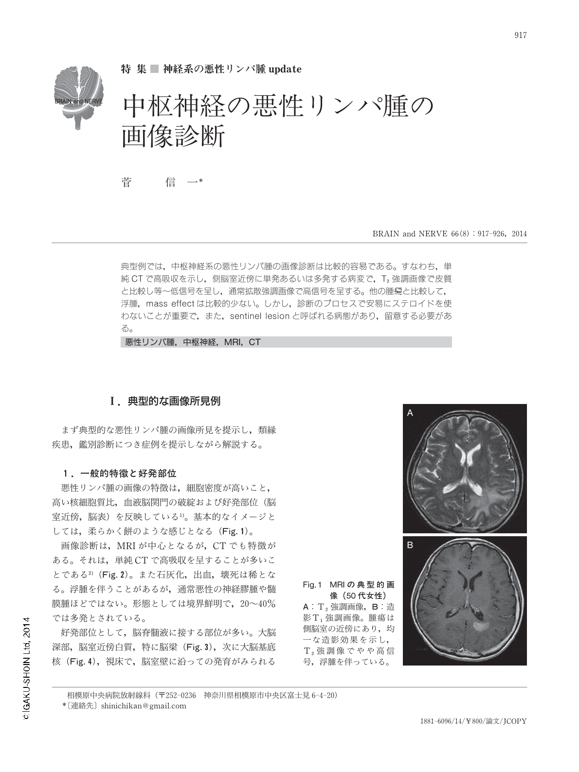
典型例では,中枢神経系の悪性リンパ腫の画像診断は比較的容易である。すなわち,単純CTで高吸収を示し,側脳室近傍に単発あるいは多発する病変で,T2強調画像で皮質と比較し等~低信号を呈し,通常拡散強調画像で高信号を呈する。他の腫瘍と比較して,浮腫,mass effectは比較的少ない。しかし,診断のプロセスで安易にステロイドを使わないことが重要で,また,sentinel lesionと呼ばれる病態があり,留意する必要がある。
Abstract
With a typical case, imaging diagnosis of central nervous system malignant lymphoma is not difficult. High density on non contrast CT, periventricular location, homogenous contrast enhancement, iso- to hypointensity to gray matter on T2 weighted MR imaging and high intensity on diffusion weighted MR imaging are characteristic findings. Hemorrhage is rare. When a patient is immunocompromised, irregular ring enhancement is noted on enhanced study.
Intravascular lymphomatois is a rare type of lymphoma. A variety of imaging findings are reported.
Differential diagnosis are many. Most difficult to distinguish is a tumefactive multiple sclerosis. Most of the reported cases of tumefactive multiple sclerosis are diagnosed by brain biopsy when the brain tumor, especially malignant lymphoma is suspected.
CLIPPERS (chronic lymphocytic inflammation with pontine perivascular enhancement responsive to steroids) has been recently identified. However, there still remains whether CLIPPERS is an actual new disease entity or represents overlapping disease.

Copyright © 2014, Igaku-Shoin Ltd. All rights reserved.


