Japanese
English
- 有料閲覧
- Abstract 文献概要
- 1ページ目 Look Inside
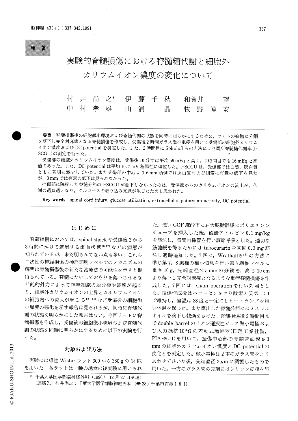
脊髄損傷後の細胞微小環境および脊髄代謝の状態を同時に明らかにするために,ラットの脊髄に分銅を落下し完全対麻痺となる脊髄損傷を作成し,受傷後2時間ガラス微小電極を用いて受傷部の細胞外カリウムイオン濃度およびDC potentialを測定した。また,2時間目にSokoloffらの方法により局所脊髄糖代謝率(1—SCGU)の測定を行った。
受傷部の細胞外カリウムイオン濃度は,受傷後10分では平均59mEqと高く,2時間目でも16mEqと高値であった。また,DC potentialは平均10.7mV程陽性に偏位した。1—SCGUは,受傷部では白質,灰白質ともに著明に減少していた。また受傷部の中心より6mm頭側では灰白質および側索に有意の低下を見たが,3mmでは有意の低下は見られなかった。
挫傷部に隣接した脊髄分節の1—SCGUが低下しなかったのは,受傷部からのカリウムイオンの流出が,代謝の過負過となり,グルコースの取り込み亢進が生じたためと思われた。
Spinal microenvironment and metabolic altera-tions after experimental contusional injury of the spinal cord were evaluated in the same Wistar rats. Severe spinal cord injury was made under light GOF anesthesia with a 10g weight drop onto the exposed Th-8 spinal cord from a 10 cm height and then halothane was ceased. The author studied extracel-lular potassium activity ([K+]e) and DC potential for 2 hours after paraplegic spinal cord injury inconscious rats. Furthermore, at 2 hours after cord injury, local spinal cord glucose utilization (1-SCGU) was measured with quantitative autoradio-grapic 2-[14C] deoxy-glucose method (Sokoloff et al.).
[K+]e) in injured spinal cords was 59±5 (mean± S. E. M.) mEq at 10 min after injury and was clear-ed with an exponential half-life of 1 hour. At 2 hours after injury [K+]e) was still high with a value of 16±1 mEq compared with 4 mEq of control animals. DC potential changes was a mirror image of that of [K+]e). DC potential changed by a mean of 10.7 mV positively from 10 min. to 2 hours after injury. 1-SCGU at theimpact site was extremely low in both white and gray matters. At 6 mm rostral from the impact center 1-SCGU was remarkably reduced in the gray matter, and in the lateral white matter. But at 3 mm rostral 1-SCGU was well preserved. And at 20 mm rostmal there was no difference in 1-SCGU with control animals.
Massive potassium efflux from the injured spinal cord to the adjacent spinal segment was clarified at this experiment. Even at 2 hours after spinal cord injury [K+]e was still high. This excess of potas-sium ion could not be buffered with only a passive glial spatial buffering mechanism, therefore it must be metabolic overload for ionic pump or other active buffering mechanisms. Relative enhance-ment of the glucose uptake at 3 mm rostral from the impact center could be due to this metabolic over-load and indicates neuronal damage.

Copyright © 1991, Igaku-Shoin Ltd. All rights reserved.


