Japanese
English
- 有料閲覧
- Abstract 文献概要
- 1ページ目 Look Inside
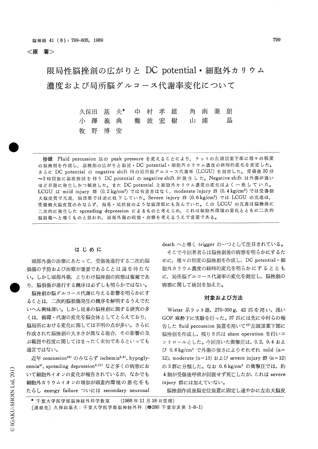
抄録 Fluid percussion法のpeak pressureを変えることにより,ラットの左頭頂葉下部に種々の程度の脳挫創を作成し,脳挫創の広がりと脳波・DC potentia1・細胞外カリウム濃度の経時的変化を測定した。さらにDC potentialのnegative shift時の局所脳グルコース代謝率(LCGU)を測定した。受傷後30分〜3時間後に脳波抑制を伴うDC potentialのnegative shiftが発生した。Negative shiftは外傷が強いほど早期に発生しかつ頻発した。またDC potentialと細胞外カリウム濃度の変化はよく一致していた。LCGUはmild injury群(0.2kg/cm2)では有意差はなく,moderate injury群(0.4kg/cm2)では受傷側大脳皮質で亢進,脳深部では逆に低下していた。Severe injury群(0.6kg/cm2)ではLCGUの亢進は,受傷側大脳皮質のみならず,海馬・尾状核のような脳深部にも及んでいた。このLCGUの亢進は脳挫創に二次的に発生したspreading depressionによるものと考えられ,これは細胞外環境の悪化とともに二次的脳損傷へと導くものと思われ,頭部外傷の病態・治療を考えるうえで重要である。
Pathophysiology of the traumatized brain, espe-cially that of cerebral contusion, is very complex and has not been well understood. In recent years, changes in extracellular ion concentration have been known in various pathological conditions such as cerebral concussion, spinal contusion, ischemia, hypoglycemia, epilepsy and spreading depression as one of the triggers to lead to secondary brain damage. To know the metabolic and ionic chan-ges following cerebral contusion, the authors made various degree of cerebral contusion by fluid per-cussion method, and observed successive changes in EEG, DC potential, extracellular potassium con-centration and local cerebral glucose utilization (LCGU).
Materials and Methods: Using 42 male Wistar rats, mild (0. 2 kg/cm2), moderate (0. 4 kg/cm2) and severe contusion (0. 6 kg/cm') were made in the left lower parietal region of the rats. EEG, DC poten-tial and extracellular potassium concentration (us-ing potassium sensitive glass microelectrode) were monitored for four to five hours after making the contusions. LCGU (by '4C-2-deoxyglucose method) was studied at the time of the negative shift of DC potential.
Results: The negative shift of DC potential with EEG supression was observed at 30 min. to 3 hours after injury. The severer the injury was, the earli-er and the more frequent negative shifts appear-ed. LCGU showed no significant changes in the mild injury group. In the moderate injury group, frequent negative shifts of DC potential associated with EEG supression were observed. A 20% in-crease of glucose utilization in the cortex of thelesion side was observed whereas 50% decreases in the subcortical structures were found. In the severe injury group, EEG was supressed immedia-tely after contusion and had never recovered. DC potential fluctuated and was unstable. The increase of LCGU was noted not only in the cortex of the lesion side but also in some of the subcortical structures (hippocampus, caudate nucleus, dentate nucleus and thalamus). The extracellular potassium concentration rose to 30 mM, being correlatedclosely with DC potential.
Discussion : Increase of LCGU associated with EEG supression, negative shift of DC potential and elevation in extracellular potassium concentration was thought to be due to spreading depression. It was postulated that spreading depression following cerebral contusion causes energy failure and can lead to secondary brain damage.

Copyright © 1989, Igaku-Shoin Ltd. All rights reserved.


