Japanese
English
- 有料閲覧
- Abstract 文献概要
- 1ページ目 Look Inside
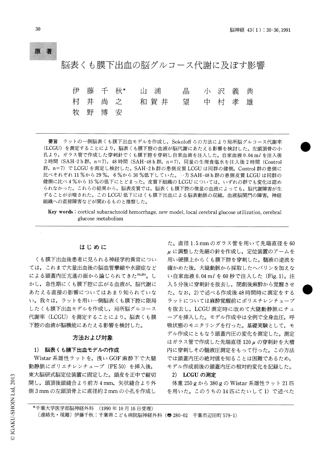
ラットの一側脳表くも膜下出血モデルを作成し,Sokoloffらの方法により局所脳グルコース代謝率(LCGU)を測定することにより,脳表くも膜下腔の血液が脳代謝にあたえる影響を検討した。左頭頂骨の小孔より,ガラス管で作成した穿刺針でくも膜下腔を穿刺し自家血液を注入した。自家血液0.04mlを注入後2時間(SAH−2 h群,n=7),48時間(SAH−48 h群,n=7),同量の生理食塩水を注入後2時間(Control群,n=7)でLCGUを測定し検討した。SAH−2 h群の患側皮質LCGUは同群の健側,Control群の患側に比べそれぞれ11%から29%,6%から30%低下していた。一方SAH−48 h群の患側皮質LCGUは同群の健側に比べ4%から15%の低下にとどまった。皮質下組織のLCGUについては,いずれの群でも変化は認められなかった。これらの結果から,脳表皮質では,脳表くも膜下腔の微量の血液によっても,脳代謝障害が生ずることが示唆された。このLCGU低下にはくも膜下出血による脳表動脈の収縮,血液脳関門の障害,神経組織への直接障害などが関わるものと推察した。
Neurological deficits following human subarach-noid hemorrhage (SAH) have been related to the cerebral arterial spasm and the increase in intra-cranial pressure (ICP) secondary to the devolop-ment of hydrocephalus. Metabolic depression in experiment study was thought as resulting from brain stem dysfunction. On the other hand, some reports have shown no relationship between vasospasm and neurological abnormalities. The mechanism of cerebral metabolism depression after SAH remains unclear. The effect of blood in the cortical subarachnoid space on the crebral metabo-lism has not been known well. To investigate this effect, a new cortical SAH model was developed using rat and local cerebral glucose utilization (LCGU) after production of SAH was measured.
A cortical SAH model : a small burr hole was made on the left parietal bone and the arachnoid membrane was pierced with a tapered glass-needle 60μ tip in diameter. Fresh autologous non-heparinized arterial blood 0.04 ml was injected into subarachnoid space within 60 seconds through that needle. The blood extended over the left cerebral cortical surface with thin subarachnoid hematoma on the parietal cortex, but did not extend on the right hemisphere and the basal cistern. The increase in ICP during production of SAH was minimal, mean value of 7.2 mmHg and ICP slowly returned to the basal level within 30 minutes.
Rats were divided into 3 groups ; rats 2 hours (SAH-2h, n=7) and 48 hours (SAH-48h, n=7) after production of SAH and rats 2hours after 0.04ml saline injection for control (Control, n=7). LCGU was studied according to the methods devel-oped by Sokoloff.
In SAH-2h, cortical LCGU on the affected side was significantly reduced compared with the non-affected side of the same group and with the affected side of Control by 11 % to 29 % and 6 % to 30 % on the average, respectively. In SAH-48h, reduction of cortical LCGU of the affected side was 4 % to 16 % and 3 % to 20 % on the average respec-tively, which was smaller than that in SAH-2h but not significant. LCGU of the subcortical structures in any groups was unchanged.
These results show that the blood in the subarach-noid space itself decreases the cortical glucose metabolism and imply that it will bring about cere-bral dysfunction. Some reports refer to the effects of the blood in the subarachnoid space on cortical structures ; constriction of pial and intraparen-chymal arteries and arterioles, breakdown of blood-brain-barier and direct damage to neurons and glial cells. The mechanism of decrease in LCGU could not be made clear, but decrease seems to reflect cortical dysfunction following these effects of subarachnoid blood.

Copyright © 1991, Igaku-Shoin Ltd. All rights reserved.


