Japanese
English
- 有料閲覧
- Abstract 文献概要
- 1ページ目 Look Inside
I.はじめに
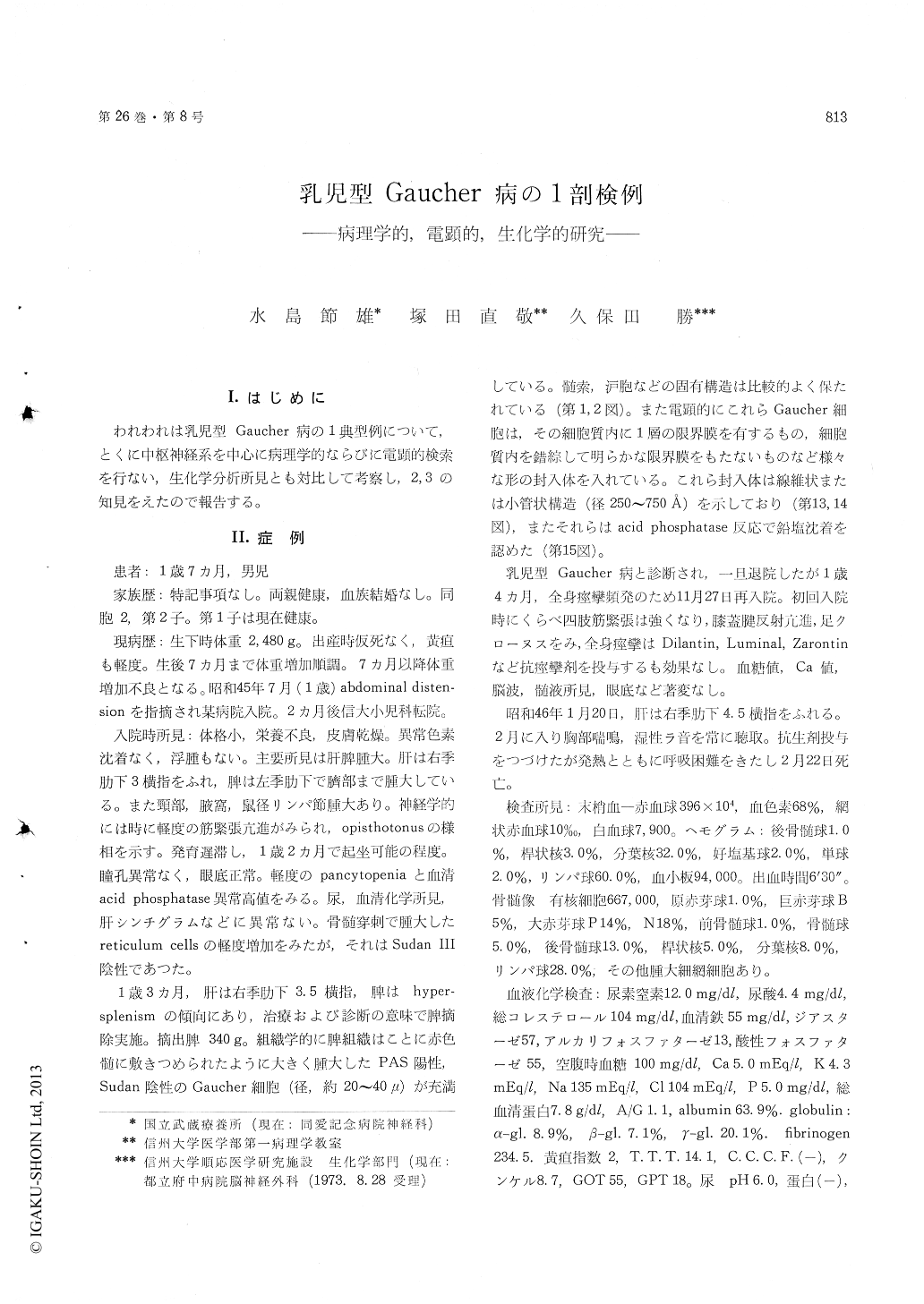
われわれは乳児型Gaucher病の1典型例について,とくに中枢神経系を中心に病理学的ならびに電顕的検索を行ない,生化学分析所見とも対比して考察し,2,3の知見をえたので報告する。
A case of infantile Gaucher's disease was ex-amined pathologically, electronmicroscopically and biochemically.
This male infant died at 19 months. He was the second child of unrelated and healthy parents and his brother is healthy now.
The patient developed normally until 7 months. At 14 months he was taken to Medical Hospital of Shinshu University because of poor development and abdominal distension. On admission, hepato-splenomegaly and marked enlargement of cervical, axillary and inguinal lymph nodes were noted. After 14 months he could no longer sit and his neurological development began to regress. Hesometimes showed hyperextension of the head, convulsion attacks and increased patellar tendon reflexes.
Laboratory tests revealed fairly pancytopenia and marked elevated serum acid phosphatase. At 16 months the spleen was removed surgically because of a remarkable sign of hypersplenism. The spleen weighed 340g, and appeared a large number of Gaucher cells which contained PAS positive and Sudan negative substances. Also, numerous spindle-shaped inclusion bodies were found in these cells under electronmicroscope. At 19 months he died of acute bronchopneumonia.
Autopsy revealed bilateral bronchopneumonia and enlargement of the liver. Histologically, in the reticuloendothelical systems (liver, bone marrow, lymph nodes, thymus, adrenals and lungs) appeared a number of Gaucher cells.
Neuropathologically, there was wide spread of acute degenerative changes in the nervous system and the lesions of greatest severity in the dentate nuclei, thalamus, cerebral cortex (parietal, insular, cingulate, occipital and frontal gyrus) and basal ganglia. Small groups of swollen nerve cells were present in the hippocampus, thalamus, basal ganglia, dentate nuclei and cerebral cortex. Proliferation of astrocytos and microglia within the 3-5 layers of the cerebral cortex was observed. Vascular changes with giant cells (Gauchercells) were found in the cerebral and cerebellar cortex, subcortical white matter and thalamus. The Gaucher cells containing spindle-shaped inclusion bodies were also found in these areas electron-microscopically. Heterotopic neurons were present in the white matter of frontal and temporal regions.
Intracytoplasmic inclusions were observed in pro-bable neurons electronmicroscopically. There were 3 types of inclusion : 1) membranous cytoplasmic bodies (MCB). -cornu Ammonis-2) granulo-membranous bodies. -dentate nuclei-3) membran-ous cytosomes filled with flat or finger print like parallel membranes. -cerebellar cortex-
Biochemically, a remarkable increase in concen-tration of glucocerebroside (glc-cer) was found in the reticuloendothelial systems. It was also found that the glc-cer increased slightly in brain and oc-cupied about 16% of kerasin fraction. A fairly amount of GM2 ganglioside was also found in this case. The cerebral glc-cer and GM2 ganglioside were composed of C 18:0 stearic acid (88.8%, 87.9%) as major fatty acid component. On the other hand, the visceral glc-cer storaged in reticulo-endothelial systems was composed of C 16:0, C 18:0, C 24:0 and C 24:1 as main fatty acid. The major glycolipid of erythrocytes was globoside-I and contained both normal and hydroxy fatty acids.

Copyright © 1974, Igaku-Shoin Ltd. All rights reserved.


