Japanese
English
- 有料閲覧
- Abstract 文献概要
- 1ページ目 Look Inside
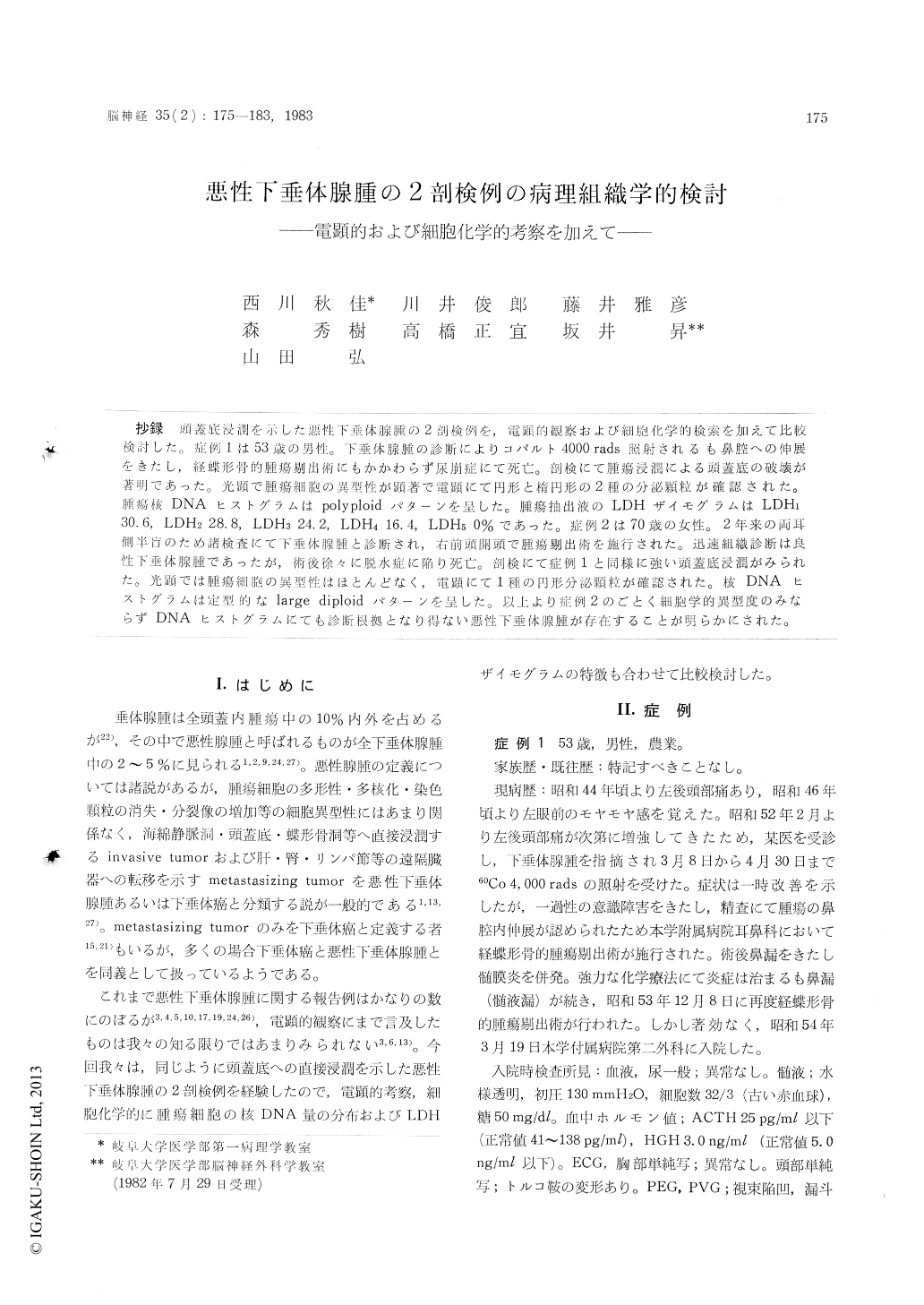
抄録 頭蓋底浸潤を示した悪性下垂体腺腫の2剖検例を,電顕的観察および細胞化学的検索を加えて比較検討した。症例1は53歳の男性。下垂体腺腫の診断によりコバルト4000rads照射されるも鼻腔への伸展をきたし,経蝶形骨的腫瘍剔出術にもかかわらず尿崩症にて死亡。剖検にて腫瘍浸潤による頭蓋底の破壊が著明であった。光顕で腫瘍細胞の異型性が顕著で電顕にて円形と楕円形の2種の分泌顆粒が確認された。腫瘍核DNAヒストグラムはpolyploidパターンを呈した。腫瘍抽出液のLDHザイモグラムはLDH130.6,LDH228.8,LDH324.2,LDH416.4,LDH50%であった。症例2は70歳の女性。2年来の両耳側半盲のため諸検査にて下唾体腺腫と診断され,右前頭開頭で腫瘍剔出術を施行された。迅速組織診断は良性直下垂体腺腫であったが,術後徐々に脱水症に陥り死亡。剖検にて症例1と同様に強い頭蓋底浸潤がみられた。光顕では腫瘍細胞の異型性はほとんどなく,電顕にて1種の円形分泌顆粒が確認された。核DNAヒストグラムは定型的なlarge diploidパターンを呈した。以上より症例2のごとく細胞学的異型度のみならずDNAヒストグラムにても診断根拠となり得ない悪性下垂体腺腫が存在することが明らかにされた。
Two autopsy cases of mailgnant pituitary ade-noma which invaded into the base of the skull are reported with some ultrastructural and cyto-chemcial characterization.
Case 1. The 53-year-old man had complained of left occipitalgia for about 2 years before he was diagnosed of pituitary tumor. He was treated with 60Co irradiation up to 4,000 rads. However, the tumor extended to the nasal cavities and the patient developed severe diabetes insipidus and dysequilibrium of electrolytes and died in-spite of the surgery for tumor resection by trans-sphenoidal approach. At autopsy, the base of the skull was destroyed with invasion of the tumor. Microscopically, the tumor tissue was composed of the cells with marked nuclear atypism. Ultrastructurally, two types of secretory granules were seen. One was in round shape and 100-250nm sized and the other one had elliptical form with 100-150 x 200-500 nm size. Nuclear DNA histogram of the tumor cells disclosed a polyploid pattern whi h suggested a malignant nature of the tumor. LDH isoenzyme proportion of the extracted tumor tissue was LDH1-30.6%, LDH2-28.8%, LDH3-24.2%, LDH4-16. 4% and LDH5-0%.
Case 2. The 70-year-old woman has complain-ed of bitemporal hemianopsia before admission to Gifu University Hospital. On the admission, she was suspected of pituitary tumor on the X-ray examination indicating calcification in the pituitary region and deformity of the sella turcica. Histological diagnosis of the tumor with the material from the surgery was benign chromo-phobe adenoma. Six months after the operation, she died of dehydration and edema. At autopsy,marked destruction of the base of the skull was noted also in this case. Microscopically, the tumor cells contained relatively uniform nuclei without prominent atypism. Ultrastructural analysis clari-fied the existence of secretory granules with round shape and 100 nm size in the cytoplasm. Nuclear DNA histogram indicated a large diploid distribution suggestive of benign tumor.
Thus, case 1 was considered to be a pituitary carcinoma with the cytochemical as well as morphological characteristics. However, case 2 in the present study revealed no cytomorphological and cytochemical evidences of malignancy. The latter case suggests to show a possibility of dis-agreement between these markers and the biologi-cal property like invasiveness of the tumor cells.

Copyright © 1983, Igaku-Shoin Ltd. All rights reserved.


