Japanese
English
- 有料閲覧
- Abstract 文献概要
- 1ページ目 Look Inside
はじめに
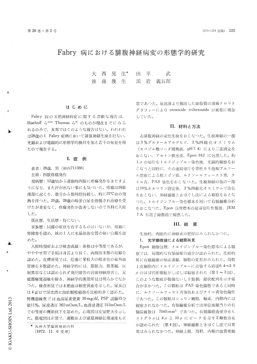
Fabry病の末梢神経病変に関する詳細な報告は,Bischoffら1)2)Thomasら3)のものが現在までにみられるのみで,本邦ではこのような報告はない。われわれは28歳の1 Fabry症例において腓腹神経生検を行ない,光顕および電顕的に形態学的検討を加え若干の知見を得たので報告する。
Light and electronmicroscopic examinations of a biopsied sural nerve from a 28-year-old patient who suffered from the deep pains in both limbs and a remarkable decrease of sweating on exer-cises and was diagnosed clinically as Fabry's disease, were reported.
In the toluidine blue stained Epon-sections the number of the myelinated fibers per square milli-meter was slightly decreased, but the degenerative changes of them were relatively rare. Electron-microscopically the degenerative changes of the axoplasm were relatively rare in both myelinated and unmyelinated fibers.
In the sections stained with the toluidine blue, abnormal fine granular inclusions were easily visualized in the perineurial cells. The inclusions were stained with the stain by Luxol Fast Blue and Nile Blue, and gave a positive reaction with the standard PAS technique. Electronmicroscopy demonstrated that the lipid droplets were present in the capillary endothelia and their pericytes as well as in the perineurial cells and not in Schwann cells. They were present in the epineurial fibro-blasts and not in both perineurial and endoneurial fibroblasts. They were identified as irregular but generally round myelin figures, with diameters reaching a little over 1.5μ. Many of them showed a distinct single limiting membrane. High reso-lution photographs revealed that they were made up of sheets or concentrically arranged lamellae showing a regular alteration of light and dark zones with a periodicity of approximately 42-50 Angstroms. In addition to these densely packed lamellae, the membrane-bounded structures which contain lipids or loosely packed lamellae were sometimes observed chiefly in both the capillary endothelia and perineurial cells. It could be con-sidered that the densely packed lamellae have originated from these membrane-bounded struc-tures. Because the membrane-bounded inclusion bodies were often observed in both the perineurial and endothelial cells with the dilated endoplasmic reticulum, it could be assumed that the formation of the inclusions is related to the endoplasmic reticulum. Multi Vesicular Bodies in the cytoplasm of the endothelial cells which were disclosed in this case had not been well documented.

Copyright © 1974, Igaku-Shoin Ltd. All rights reserved.


