Japanese
English
- 有料閲覧
- Abstract 文献概要
- 1ページ目 Look Inside
緒言
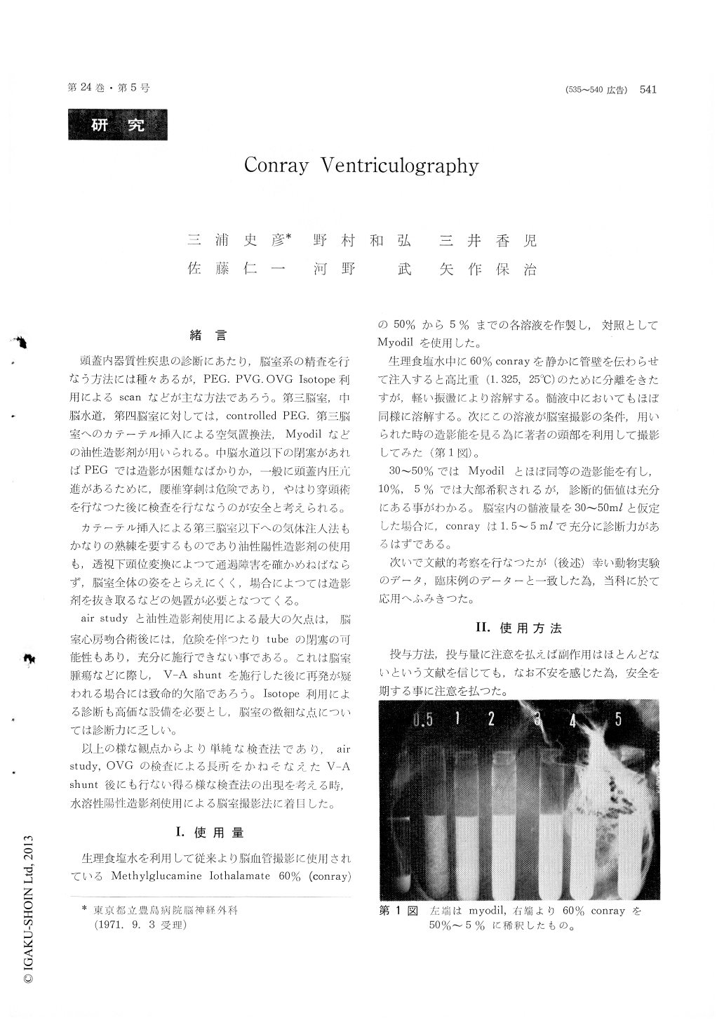
頭蓋内器質性疾患の診断にあたり,脳室系の精査を行なう方法には種々あるが,PEG.PVG.OVG Isotope利用によるscanなどが1こな方法であろう。第三脳室,中脳水道,第四脳室に対しては,controlledPEG.第三脳室へのカテーテル挿入による空気置換法,Myodilなどの油性造.影剤が用いられる。中脳水道以下の閉塞があればPEGでは造影が困難なばかりか,一般に頭蓋内圧亢進があるために,腰椎穿刺は危険であり,やはり穿頭術を行なつた後に検査を行ななうのが安全と考えられる。
カテーテル挿入による第1脳室以下への気体注入法もかなりの熟練を要するものであり油性陽性造影剤の使用も,透視下頭位変換によつて通過障害を確かめねばならず,脳室全体の姿をとらえにくく,場合によつては造影剤を抜き取るなどの処置が必要となつてくる。
This paper presents twenty-one clinical cases re-garding ventriculography with positive contrast medium conray. Ventriculographies with conray (methylglucamine isothalamate sixty percents were attempted and good radiographic appearances of the ventricular system, especially, the third ventricle, the aqueduct and the fourth ventricle were obtained, even if sixty percent conray was diluted down to five to ten percents.
Two to five mililiters of sixty percent conray was injected into the lateral ventricle. One of the cases described in this paper showed that the ven-tricle atrial shunt tube was visualized five minutes after the injection.
Side effects rarely appeared. In one of the cases, the patiant had high fever through two days. In another case, misinjection into the interhemisphric fissure developed clonic seizure and tonic spasm on the patiant's lower limbs through ninety minutes.
This method had little side effect and revealed the much clearer radiographic visualization of the ventricular system in comparison with air study or oleoventriculography.

Copyright © 1972, Igaku-Shoin Ltd. All rights reserved.


