Japanese
English
- 有料閲覧
- Abstract 文献概要
- 1ページ目 Look Inside
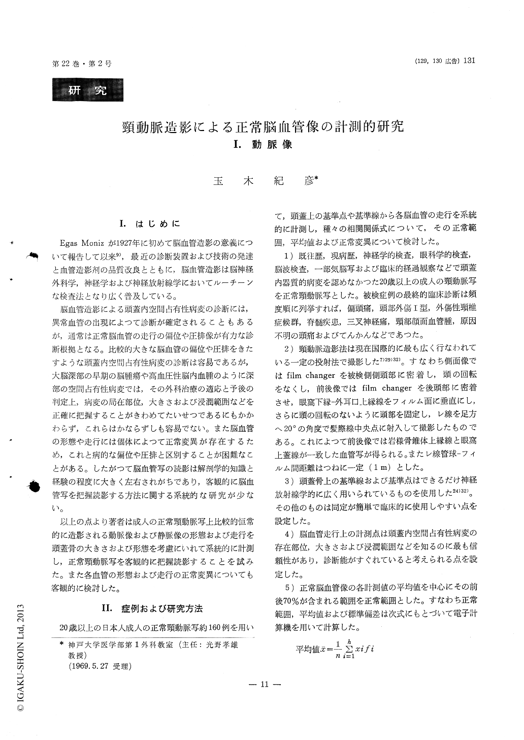
I.はじめに
Egas Monizが1927年に初めて脳血管造影の意義について報告して以来9),最近の診断装置および技術の発達と血管造影剤の品質改良とともに,脳血管造影は脳神経外科学,神経学および神経放射線学においてルーチーンな検査法となり広く普及している。
脳血管造影による頭蓋内空間占有性病変の診断には,異常血管の出現によつて診断が確定されることもあるが,通常は正常脳血管の走行の偏位や圧排像が有力な診断根拠となる。比較的大きな脳血管の偏位や圧排をきたすような頭蓋内空間占有性病変の診断は容易であるが,大脳深部の早期の脳腫瘍や高血圧性脳内血腫のように深部の空間占有性病変では,その外科治療の適応と予後の判定上,病変の局在部位,大きさおよび浸潤範囲などを正確に把握することがきわめてたいせつであるにもかかわらず,これらはかならずしも容易でない。また脳血管の形態や走行には個体によつて正常変異が存在するため,これと病的な偏位や圧排と区別することが困難なことがある。したがつて脳血管写の読影は解剖学的知識と経験の程度に大きく左右されがちであり,客観的に脳血管写を把握読影する方法に関する系統的な研究が少ない。
Topometric study of the normal carotid arte-riograms was made, using about 160 normal carotid arteriograms which had come from only the pa-tients over the ages of twenty in whom the cerebral organic lesions were ruled out in history, neurologic and ophthalmic examination, electroencephalographic examination, cerebrospinal fluid study, pneumoence-phalography and subsequent follow up of the clinical course.
The angiographic measurements of the position and the configuration of the anterior cerebral artery, middle cerebral artery, lenticulostriate arteries and anterior choroid artery were statistically analysed in both the lateral and the antero-posterior plane.
The present study established the normal limits of the position and configuration of the various por-tions of the each arteries. Normal variation of the each arteries was also statistically analysed and des-cribed.
1. Anterior cerebral artery :
The cranial landmarks and basic lines for measure-ment were illustrated in Figure 1 in the lateral and Figure 2 in the antero-posterior projections. Lateral view ; The distance from the fronto-basal point of the anterior cerebral artery to the nasion was 33.2± 2.6% of the distance from the nasion to the en-dolambda. The distance from the fronto-polar point of the anterior cerebral artery to the point on the frontal inner table of the skull 4 cm over the nasion was 23.7±2.9% of the distance from the the point on the fronal inner table to the endolambda.
The distance from the parietal point of the an-terior cerebral artery to the endobregma was 36.5 ± 3.3% of the distance from the endobregma to the external auditory meatus. The distance from the parieto-occipital point of the anterior cerebral artery to the inner table of the skull in the parieto-occipital region to the external auditory meatus.
The parietal portion of the anterior cerebral artery was subjected to the more variations than the other portion of its artery.

Copyright © 1970, Igaku-Shoin Ltd. All rights reserved.


