Japanese
English
- 有料閲覧
- Abstract 文献概要
- 1ページ目 Look Inside
近年,胃癌の診断については内視鏡およびX線検査の急速な進歩により,微細病変の診断まで可能となった.しかしこれらの方法は胃壁を粘膜側から検査する方法であり,癌の深達度および浸潤範囲を誤診する場合が少なくない.そこで動脈造影を補助診断として用いることにより正確な深達度,浸潤範囲を読みとることを試みた.さらに動脈造影上の血管所見と腫瘍血管がどのような関連をもっているかを解明すべく,微細血管構築像の手技を用いて,両者の血管所見を分析し検討した.
Recently the angiographic diagnostic method has made a remarkable progress as an effective method of identifying cancers of various organs. However, there were few discussions concerning the studies of angio-graphic vascular patters in the tumor vessels. In order to elucidate this problem, we have selected gastric cancer because of its extremely variegated histology and morphology. Utilizing Seldinger techniques, we have carried out comparative studies between the superselective angiographical patterns, especially of the left gastric artery, and microangiographical pictures of the resected specimen in the same patients.
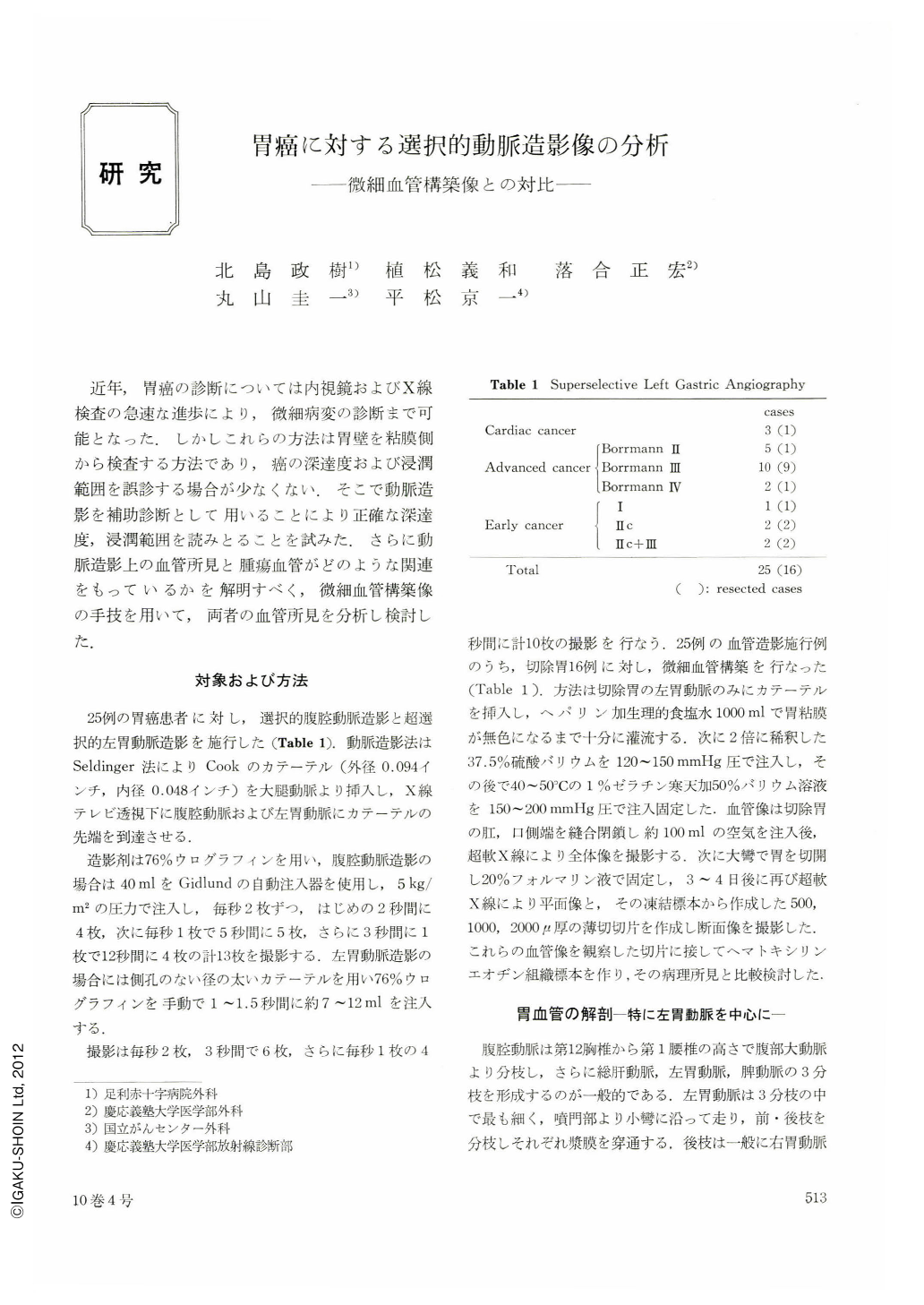
Superselective angiography was performed exclusively on 3 patients with cardiac cancer, 17 patients with advanced cancer (5 cases Borrmann Ⅱ, 10 of Borrman Ⅲ and 2 of Borrman IV) and 5 patients with early cancer (1 case of type Ⅰ, 2 each of Ⅱc and Ⅱc+Ⅲ).
Classification of the vascular patterns of tumors based on the angiographic pictures was (1) encasement, (2) vascular occlusion, (3) neovascularity, (4) tumor stain, (5) venous laking (lake) and (6) venous drainage. On the other hand, microangiography revealed that vascular pattern of gastric cancer, different from that of the normal stomach, showed special vascular architectures. Besides, the vascular pattern varies according to the types of lesions, degrees of invasion and histopathologic findings as follows:
1. Seen from the degree of vascular density, cancers are classified into hypervascular and hypovascular types.
2. The tumor vessels are tortuous, narrow or straight according to the histology of the tumors. Although differences exist in the level of the contrast medium between angiography and microangiography, comparative studies show that encasement represent direct infiltration into the vessels of the subserous and submucosal layers. Vascular occlusion results from the isolation and congestion of the same vessels. Neovascularity shows tumor vessels of hypervascular cancer with congestion in the borders of the tumor. In the case of tumor stain, findings are almost similar to those of neovascularity. Venous laking is interpreted as congestion of the borders of the tumor, while venous drainage is presumed to be due to the formation an arterio-venous fistula with descructive development of cancer at the borders of the tumor.
Copyright © 1975, Igaku-Shoin Ltd. All rights reserved.