Japanese
English
- 有料閲覧
- Abstract 文献概要
- 1ページ目 Look Inside
I.緒言
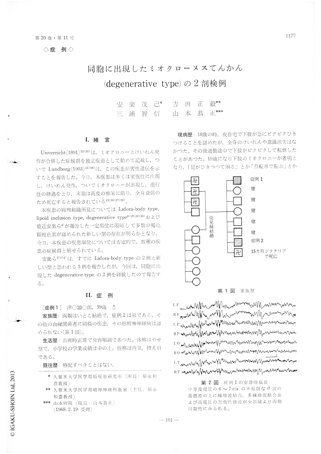
Unverricht (1891)25)26)は,ミオクロニーとけいれん発作が合併した症候群を独立疾患として始めて記載し,ついでLundborg (1903)15)16)は,この疾患が劣性遣伝を示すことを報告した。今日,本疾患は多くは家族性に出現し,けいれん発作,ついでミオクロニーが出現し,進行性の経過をとり,末期は高度の痴呆に陥り,全身衰弱のため死亡すると報告されている19)20)27)30)。
本疾患の病理組織所見についてはLafora-body type, lipoid inclusion type, degenerative type8)19)20)30)および最近安楽ら4)が報告した一定部位に限局して多数の褐色顆粒色素が認められた新しい型の存在が明らかとなり,今日,本疾患の疾患単位については否定的で,数種の疾患の症候群と解せられている。
Case 1. A 39-year-old man, whose parents were cusins, was attacked with myoclonia at the age of 18. Two years later grand mal came to appear and then cerebellar symptoms, dementia and changes of personality gradually became noticeable. The pati-ent died of marasmus 21 years after the onset ofthe disease.
Histopathological findings of the brain :
Nerve cells in the dentate nuclei of the cerebellum were generally atrophic, Purkinje cells were parti-ally atrophic, and so were nerve cells in the olivary nuclei. Nerve cells in the cerebral cortex presented atrophy and shadow figures along with a disorder in the cytoarchitecture in the frontal lobe. What is more interesting is that the hippocampus major showed, though slightly, an Alzheimer's fibrillary degeneration and a granulovacuolar one.
Case 2. A man aged 26, a younger brother of Case 1, was also attacked with myoclonia at the age of 15. Having taken the same course as in Case 1,the patient died of hemorrhagic pneumonia after the lapse of 11 years.
Histopathological findings of the brain :
Nerve cells in the dentate nuclei of the cerebellum presented an advanced atrophy and a marked de-position of lipofuscin. In the dentate nuclei, the proliferation of glial cells was also noted. Most of Purkinje cells fell off diffusely and the remainder were atrophic. A part of granular cells were also pyknotic and irregular in shape. The changes observed in the olivary nuclei and the cerebral cor-tex were almost the same those in Case 1.

Copyright © 1968, Igaku-Shoin Ltd. All rights reserved.


