Japanese
English
- 有料閲覧
- Abstract 文献概要
- 1ページ目 Look Inside
I.はじめに
眼窩静脈撮影は眼窩内の血管腫など特異な疾患の診断にきわめて有効な方法とされているが,従来施行されてきたのはV. angularis1)〜3)穿刺による方法でありその手技はなかなか困難であり,失敗すると大きな血腫を内眥部に作つたりするため敬遠されがちであつた。
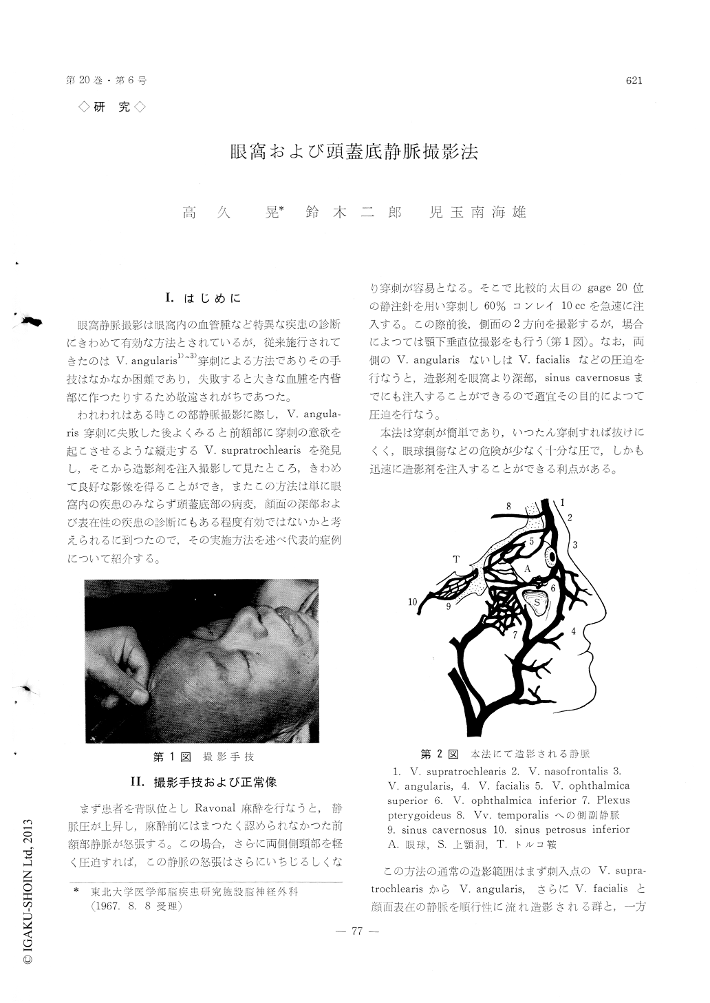
われわれはある時この部静脈撮影に際し,V. angula—ris穿刺に失敗した後よくみると前額部に穿刺の意欲を起こさせるような縦走するV. supratrochlearisを発見し,そこから造影剤を注入撮影して見たところ,きわめて良好な影像を得ることができ,またこの方法は単に眼窩内の疾患のみならず頭蓋底部の病変,顔面の深部および表在性の疾患の診断にもある程度有効ではないかと考えられるに到つたので,その実施方法を述べ代表的症例について紹介する。
A method of orbital and cavernous sinus Veno-graphy via frontal vein was reported.
The procedure of this method is more easier, safer and surer than that of the method via angular vein reported by Krayenbuhl. Not only the orbital vein but also the deep facial vein and the cavernous sinus can be visualized simultaneously by this method.
The shadow of anterior half of the cavernous sinus is visualized more clearly than the method by cateterization through into the internal jugular vein reported by Hanafee, but the shadow of the post-erior half of the cavernous sinus is inferior to the shadow of Hanafee.
This examination will aid in the diagnosis of dis-eases of the orbita, the cranial base and the nasal sinus.

Copyright © 1968, Igaku-Shoin Ltd. All rights reserved.


