Japanese
English
- 有料閲覧
PPV購入案内
1,320円 (1,200円+税10%) こちらは、2167ページから2179ページの文献です。購入には医書.jp本会員登録が必要です。
会員登録完了後に改めて上記「購入サイトへ」を選択してください。
または「購入サイトへ」選択後に「購入ログイン」、「新規会員登録」の順に進んでください。
会員登録について詳しくはをご確認ください。
- Abstract 文献概要
- 1ページ目 Look Inside
緒言
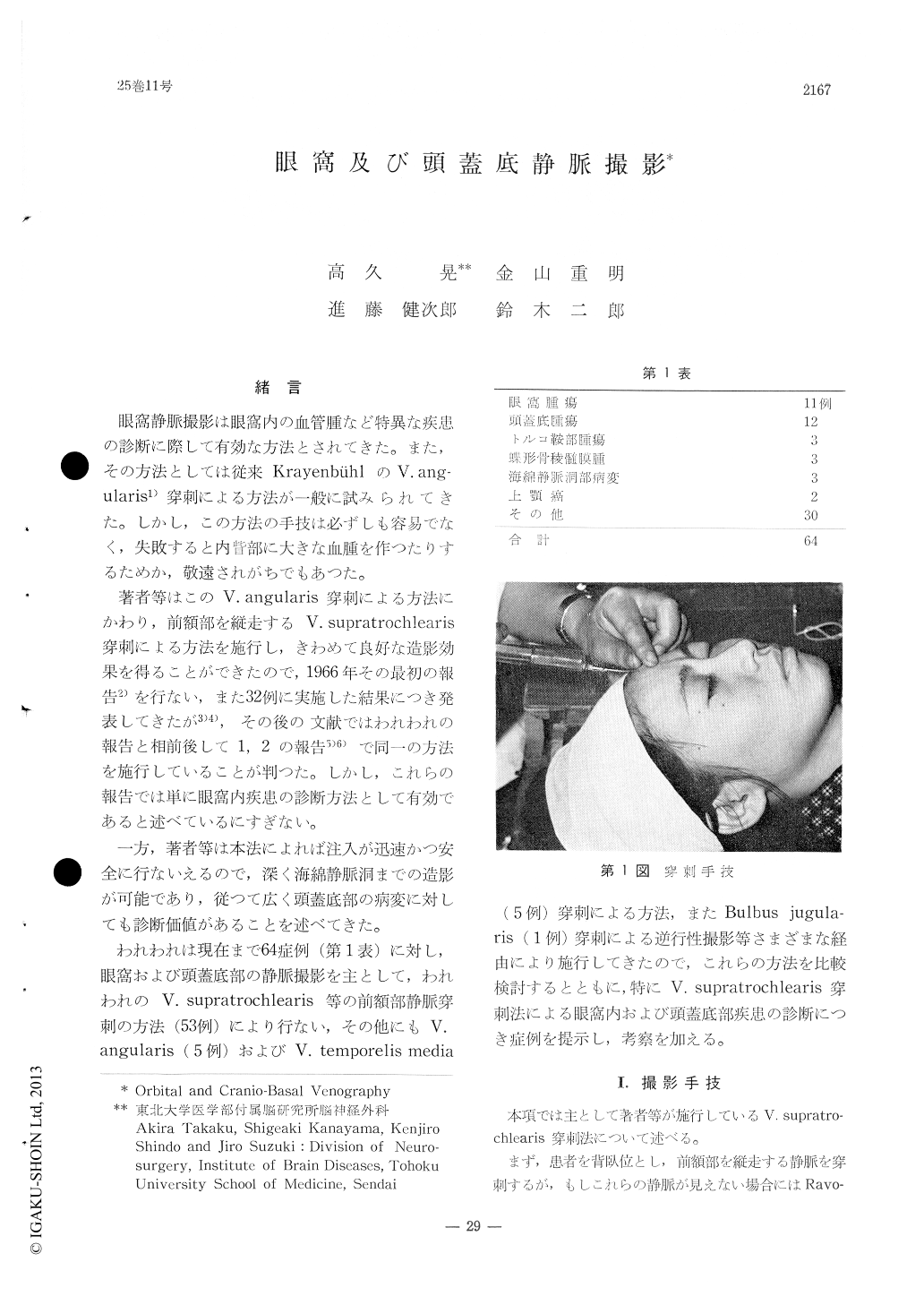
眼窩静脈撮影は眼窩内の血管腫など特異な疾患の診断に際して有効な方法とされてきた。また,その方法としては従来KrayenbühlのV.ang—ularis1)穿刺による方法が一般に試みられてきた。しかし,この方法の手技は必ずしも容易でなく,失敗すると内眥部に大きな血腫を作つたりするためか,敬遠されがちでもあつた。
著者等はこのV.angularis穿刺による方法にかわり,前額部を縦走するV.supratrochlearis穿刺による方法を施行し,きわめて良好な造影効果を得ることができたので,1966年その最初の報告2)を行ない,また32例に実施した結果につき発表してきたが3)4),その後の文献ではわれわれの報告と相前後して1,2の報告5)6)で同一の方法を施行していることが判つた。しかし,これらの報告では単に眼窩内疾患の診断方法として有効であると述べているにすぎない。
The orbital and cavernous sinus venography by supratrochlear venous puncture has been re-ported in comparison with such methods as an-gular venous puncture and/or middle temporal venous puncture. Typical intraorbital and era-nio-basal venous patterns have been presented from our series of 64 cases with various kinds of diseases.
This method is easier to perform than that of angular venous puncture technique and visua-lizes well bilateral superior ophthalmic veins as well as cavernous sinus and deep facial ve-ins.
Copyright © 1971, Igaku-Shoin Ltd. All rights reserved.