Japanese
English
- 有料閲覧
- Abstract 文献概要
- 1ページ目 Look Inside
I.緒言
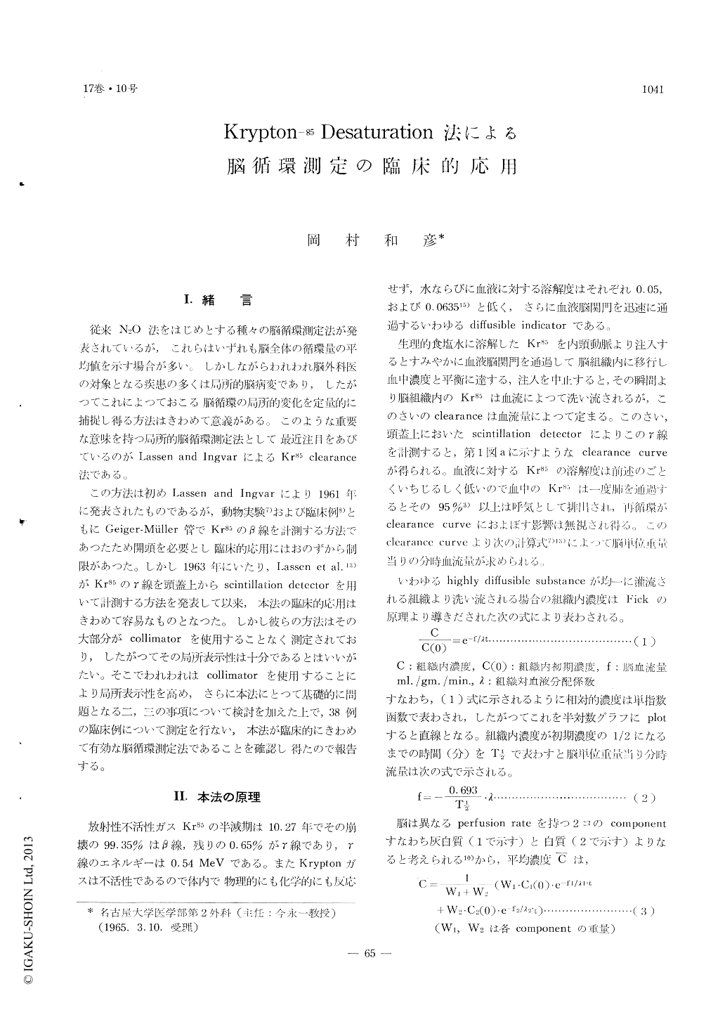
従来N2O法をはじめとする種々の脳循環測定法が発表されているが,これらはいずれも脳全体の循環量の平均値を示す場合が多い。しかしながらわれわれ脳外科医の対象となる疾患の多くは局所的脳病変であり,したがつてこれによつておこる脳循環の局所的変化を定量的に捕捉し得る方法はきわめて意義がある。このような重要な意味を持つ局所的脳循環測定法として最近注目をあびているのがLassen and IngvarによるKr85 clearance法である。
この方法は初めLassen and Ingvarにより1961年に発表されたものであるが,動物実験7)および臨床例8)ともにGeiger-Müller管でKr85のβ線を計測する方法であつたため開頭を必要とし臨床的応用にはおのずから制限があつた。しかし1963年にいたり,Lassen et al.13)がKr85のγ線を頭蓋上からscintillation detectorを用いて計測する方法を発表して以来,本法の臨床的応用はきわめて容易なものとなつた。しかし彼らの方法はその大部分がcollimatorを使用することなく測定されており,したがつてその局所表示性は十分であるとはいいがたい。そこでわれわれはcollimatorを使用することにより局所表示性を高め,さらに本法にとつて基礎的に問題となる二,三の事項について検討を加えた上で,38例の臨床例について測定を行ない,本法が臨床的にきわめて有効な脳循環測定法であることを確認し得たので報告する。
The quantitative measurements of the regional cerebral blood flow through the intact skull applying the method devised by Lassen et al. were performed on thirty-eight patients.
For the purpose of recording from a more circum-scribed area, we used a cylindrical lead collimator of 3.5cm. in diamter and 5cm. in length, and thus obtained counting of much narrower area which was confined within the limits of a lobe of the hemisphere.
The normal value of the regional cerebral blood flow (CBFr) in the frontal and the temporal region ranged from 52 to 65ml. per 100g. per minute, and from 54.0 to 71.2ml. respectively.
The mean value of CBFr in 6 patients with posttraumatic disorders was 85.2ml. in the frontal, and 37.0ml. in the temporal.
The mean value of CBFr in 6 patients with brain tumors was 33.8ml. in the temporal. These results showed that the higher the intranial pressure rose, the more severely decreased the CBFr. In a patient with approximately 200g. of angioblastic meningioma in the right parietal region, CBFr was increased over the normal value in the right temporal region in accordance with the tumor. On the contrary, it decreased severely in the opposite side probably due to the increased intracranial pressure. After removal of the tumor, CBFr restored to the normal range, namely by decreasing in the affected side and increasing in the opposite hemisphere.
In 3 patients with occlusion of the middle cerebral artery, the reduction of CBFr was evidently observed in accordance with the lesion.
Such a local change of the cerebral blood flow in these occlusive diseases would not be detected by such a method for measuring the total blood flow as N2O method by Kety and Schmidt.
The mean value of 5 patients with cerebral arterio-sclerosis was 35.4 in the frontal and 37.1ml. in the temporal region respectively.
In 4 patients with internal carotid occlusive disease, the mean value was 29.0 on affected side, and 40.2 ml. on the opposite side.
In conclusion, this method has many advantages over the other methods for measuring the cerebral blood flow. First, it can be detected a local change of the cerebral blood flow which might be missed in measurement of total cerebral blood flow as N2O method. In the second place, it can be performed very rapidly without much difficulties, and repeated measurements are possible.

Copyright © 1965, Igaku-Shoin Ltd. All rights reserved.


