Japanese
English
- 有料閲覧
- Abstract 文献概要
- 1ページ目 Look Inside
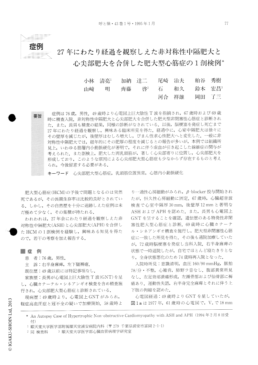
症例は76歳,男性.49歳時より心電図上巨大陰性T波を指摘され,67歳時および69歳時に精査入院.非対称性中隔肥大と心尖部肥大を合併した肥大型非閉塞性心筋症と診断された.また,長男も精査の結果,同様の診断がなされている.以後,脳梗塞を発症し死亡まで27年にわたり経過を観察し,興味ある臨床所見を得た.経過中に,心室中隔肥大は徐々にその壁厚を減じたが,後壁厚はむしろ増大し,びまん性求心性肥大へと変化した.一般に非対称性中隔肥大では,経年的にその肥厚の程度を減じるとの報告が多いが,本例では組織所見上,いわゆる筋層内小動脈硬化が著明で,それに伴う虚血が引き起こした線維症の関与が考えられた.また剖検上,肥大した両乳頭筋が,著しく心尖部寄りに位置し,心尖部肥大を形成しており,このような原因による心尖部肥大型心筋症も少なからず存在するものと考えられ,今後留意する必要がある.
We present a case of a 76-year-old male patient who, at the age of 49, was diagnosed as hypertrophic non-obstructive cardiomyopathy with both asymmetrical septal hypertrophy and apical hypertrophy. We were able to observe his condition over a period of 27 years, and get some interesting facts until the time of his death.
The thickness of the interventricular septum was reduced gradually in contrast to the increase in the inferior wall thickness. Finally, his left ventricular hypertrophic pattern became concentric. It seems that reduction of wall thickness in the interventricular se-ptum was related to progressive myocardial fibrosis caused by intramyocardial small artery sclerosis.
The ECG findings changed from high QRS voltage with giant negative T wave to normal QRS voltage with slightly negative T wave. These changes appear to have been caused by the progressive myocardial fibrosis.
The macroscopic findings of his autopsy show marked hypertrophy of the papillary muscles, which were, in turn, located extraordinarily close to the apex. This was combined with hypertrophy of the apical myocardium, which made a spade-shaped lumen with narrowing of the apical half of the left ventricle. These findings indicate that there are some cases of apical hypertrophic cardiomyopathy which are caused by the extraordinary location of the hypertrophied papillary muscles.

Copyright © 1995, Igaku-Shoin Ltd. All rights reserved.


