Japanese
English
- 有料閲覧
- Abstract 文献概要
- 1ページ目 Look Inside
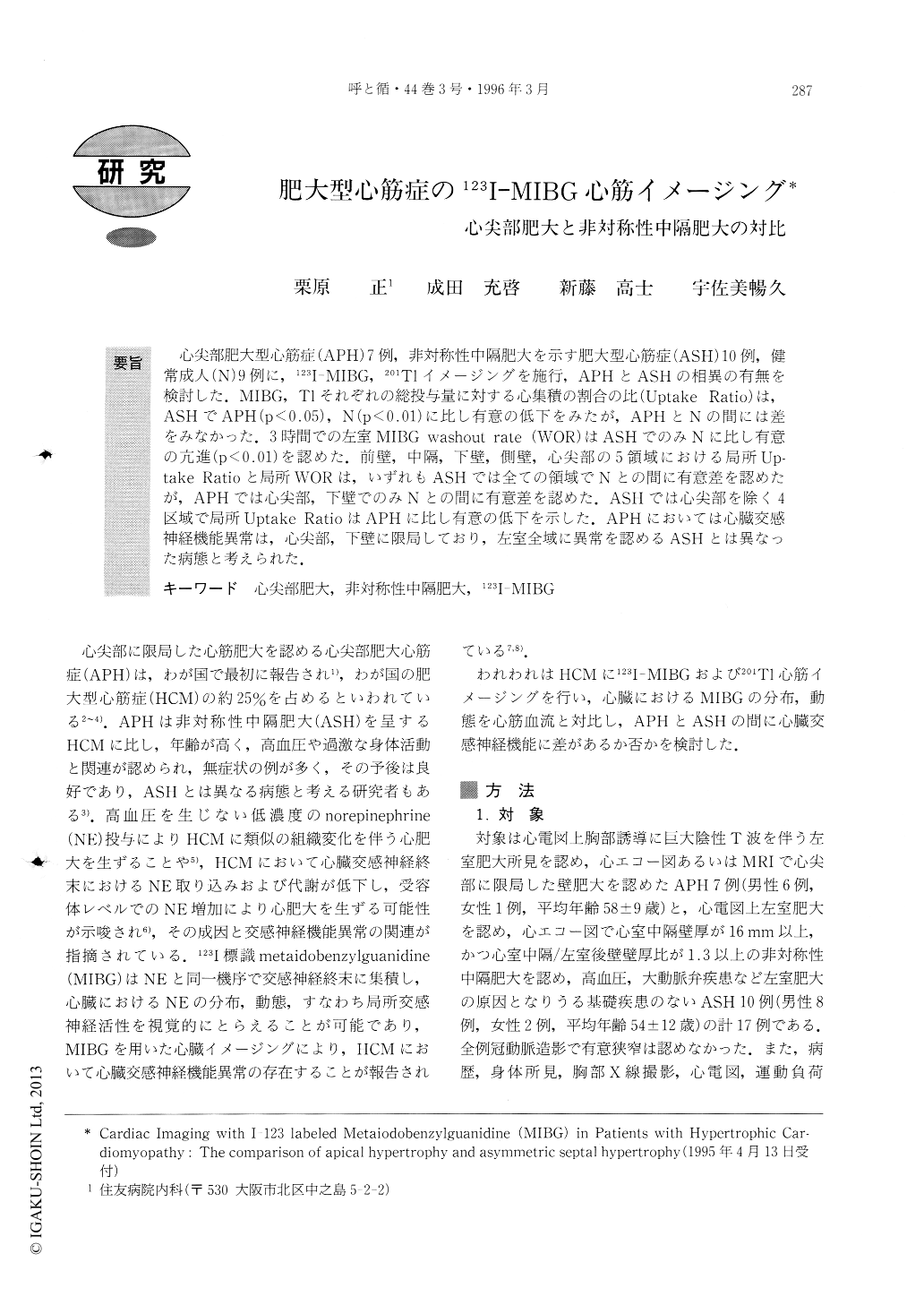
心尖部肥大型心筋症(APH)7例,非対称性中隔肥大を示す肥大型心筋症(ASH)10例,健常成人(N)9例に,123I-MDBG,201T1イメージングを施行,APHとASHの相異の有無を検討した.MIBG,T1それぞれの総投与量に対する心集積の割合の比(Uptake Ratio)は,ASHでAPH(p<0.05),N(p<0.01)に比し有意の低下をみたが,APHとNの間には差をみなかった.3時間での左室MIBG washout rate(WOR)はASHでのみNに比し有意の亢進(p<0.01)を認めた.前壁,中隔,下壁,側壁,心尖部の5領域における局所Up—take Ratioと局所WORは,いずれもASHでは全ての領域でNとの間に有意差を認めたが,APHでは心尖部,下壁でのみNとの間に有意差を認めた.ASHでは心尖部を除く4区域で局所Uptake RatioはAPHに比し有意の低下を示した.APHにおいては心臓交感神経機能異常は,心尖部,下壁に限局しており,左室全域に異常を認めるASHとは異なった病態と考えられた.
To evaluate the difference in cardiac sympathetic innervation between apical hypertrophic car-diomyopathy (APH) and hypertrophic cardiomyopathy with asymmetric septal hypertrophy (ASH), cardiac imaging with I-123 labeled metaiodobenzylguanidine (MIBG) and thallium-201 (T1) using single photon emission computed tomography and whole body scinti-graphy was performed in seven patients with APH, eleven patients with ASH and nine normal subjects. The ratio of cardiac uptake of MIBG in delayed image taken three hours after initial imaging to that of T1 in the initial image (Uptake Ratio) was significantly more reduced in ASH than in APH and normals (0.53±0.07 in ASH, 0.66±0.10 in APH; p<0.05, 0.74±0.07 in nor-mals; p< 0.01). Cardiac distribution of MIBG and T1 was homogeneous in normals, while in HCM, cardiac distribution of MIBG was different to that of T1. While increased accumulation of T1 was frequently observed in the hypertrophied region, visual defects in MIBG images were observed in the inferior to the lateral wall in ASH, and in the apex and the inferior wall in APH in more than half of the patients. Regional uptake ratio obtained in anterior, septal, inferior and lateral wall and apex were reduced more in ASH in all five regions than in normals, while in APH, regional uptake ratio was reduced in only the apex and the inferior wall, and was significantly higher than in ASH in the four regions other than the apex. Cardiac washout rate (WOR) of MIBG increased significantly more than in normals in only ASH, and there was no difference between WOR in ASH and in APH. Regional WOR increased in all five regions more in ASH than in normals, but increased more in only the apex and the inferior wall in APH than in normals. Regional WOR increased significantly more in ASH than in APH only in the anterior wall.
In conclusion, cardiac sympathetic innervation abnor-mality relative to myocardial mass was different in ASH and APH. It was localized in the region of hyper-trophy in APH, while it was observed all over the heart and was not confined to the region of hypertrophy in ASH.

Copyright © 1996, Igaku-Shoin Ltd. All rights reserved.


