Japanese
English
- 有料閲覧
- Abstract 文献概要
- 1ページ目 Look Inside
I.まえがき
Pneumographyについては,Riegel18)19)が初めて左右対称的位置で胸廓呼吸曲線を描写し,外国ではAnthony,Hansen1)2)6)13)等の報告があり,本邦に於ても石山等により種々な肺部疾患,例えば結核3)26)27),肺虚脱8)12),気管支喘息10)等及び横隔膜下膿瘍23)其他24)腹部疾患22)についての報告がある。
我々は嚢状肺気腫の病態生理の研究の途上,気腫性嚢胞が肺内の緊張性の含気嚢状物であるので,呼吸時,胸廓運動の異常が起るのでないかと考えた。この胸廓運動の測定のためにPneumo—graphを用い,又同時にBronchospirogram,Spirogramと比較検討して次の様な結果を得たので報告する。
We described a pneumogram of emphysematous bullae by means of Ishiyama-Yokoyama's pneumo-graph.
The pneumography was made symmetrically in the four points, i.e. on the mammilar line in the third, fourth intercostal spaces and on the umbilical level, and on the anterior axillar line in the ninth intercostal space.
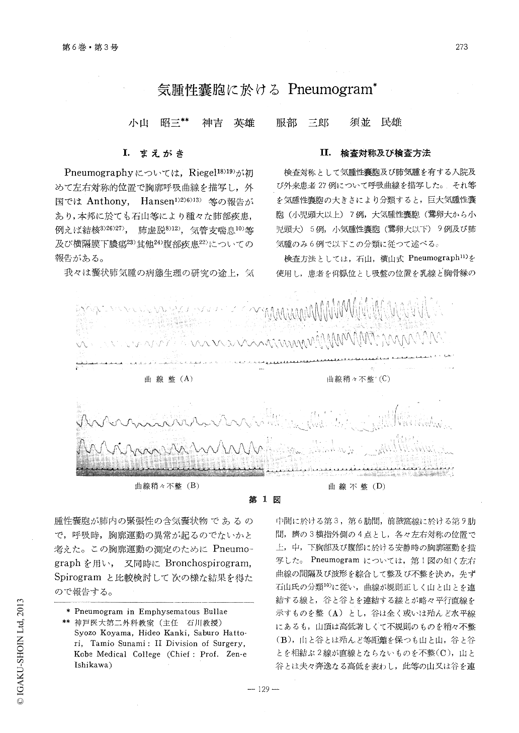
In the pneumogram we examined rate of amplitude in both sides (left and right), rhythm of the curve, prolongation of exspiration and evenness in transition portion from inspiration to exspiration. Furthermore, we investigated relation between pneumogram and grade of emphysema or broncho-spirogram (or spirgram).
We examined 27 cases of emphysematous bullae, namely giant emphysematous bullae (7 cases), large emphysematous bullae (5 cases), small emphysematous bullae (9 cases) and emphysema (6 cases).
Results :
1. In larger emphysematous bullae and advanced emphysema, the pneumogram was irregular and showed prolonged exspiration. Giant and large emphysematous bullae demonstrated distinct difference between the healthy and affected sides in amplitude of both sides (left and right) and evening in transition portion from inspiration to exspiration, expecially in the affected side.
2. In giant and large emphysematous bullae, it indicated decrease of amplitude in the affected side.
3. The pneumogram and bronchospirogram showed a positive interrelation in all except one case. In most cases, showed prolonged exspiration in pneumogram, spirogram demonstrated airtrapping.

Copyright © 1958, Igaku-Shoin Ltd. All rights reserved.


