Japanese
English
- 有料閲覧
- Abstract 文献概要
- 1ページ目 Look Inside
はじめに
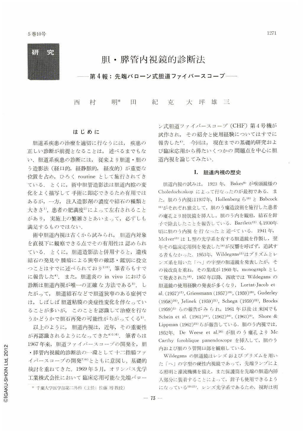
胆道系疾患の治療を適切に行なうには,疾患の正しい診断が前提となることは,述べるまでもない.胆道系疾患の診断には,従来より胆道・胆のう造影法(経口的,経静脈的,経皮的)が重要な位置を占め,ひろくroutineとして施行されてきている.とくに,術中胆管造影法は胆道内腔の変化をよく描写して手術に即応できるため有用ではあるが,一方,注入造影剤の濃度や結石の種類と大きさ1),患者の肥満度2)によって左右されることがあり,実施上の繁雑さとあいまって,必ずしも満足するものではない.
術中胆道内視は古くから試みられ,胆道内対象を直視下に観察できる点でその有用性は認められている.とくに,胆道造影法と併用すると,遺残結石の発見や腫瘍による狭窄の確認・鑑別に役立つことはすでに述べられており2)3),筆者らもすでに報告した4).また,胆道炎のin vivoにおける診断は胆道内視が唯一の正確な方法である3).したがって,胆道結石などで胆道狭窄のある症例では,しばしば胆道粘膜の炎症性変化を伴なっていることが多いが,このことを認識して治療を行なうかどうかで胆石再発の可能性がちがってくる2).
以上のように,胆道内視は,近年,その重要性が再認識されるようになってきた4)5)6).筆者らは1967年来,胆道ファイバースコープの開発を,胆・膵管内視鏡的診断法の一環として十二指腸ファイバースコープの開発7)8)とともに意図し,基礎的検討を重ねてきた.1969年5月,オリンパス光学工業株式会社において臨床応用可能な先端バローン式胆道ファイバースコープ(CHF)第4号機が試作され,その紹介と使用経験についてはすでに報告した4).今回は,現在までの基礎的研究および臨床応用から得たいくつかの問題点を中心に胆道内視を論じてみたい.
As one of series of endoscopic methods of diagnosis in diseases of the biliary and pancreatic ducts, the authors have long exploited choledochoscope along with duodenofiberscope. In this paper is introduced a fiberoptic choledochoscope manufactured for trial with a balloon equipped at its tip (CHF NO. 4-type). Its performance, the setup and direction for use is described. Endoscopic pictures of intraluminal biliary tract is also displayed as visualized by this method. The results show that this method, when combined with cho ledochography, is very effective in the diagnosis of diseases of the biliary tract.
The advantages of this method are: its simple maneuver saves time and personnel necessary for examination; removal of the bile can be done at the same time with holding a distance necessary for clear vision; and because no irrigation fluid is necessary any intra-abdominal pollution does not take place and no accumulation of fluid is seen in the lumen of the duodenum. On the other hand, it has several demerits; the reflected light of the balloon surface is superimposed on the observation field; only plane observation is possible because this method is a contact vision; it is impossible to advance the scope into the hepatic ducts with the balloon inflated as it is. Furthermore, the scope rubs against the lumen of the bile duct in removing the balloon; the balloon at the tip stands in the way of free passage of the forceps. The irrigation method hitherto employed is free from those drawbacks seen in the balloon method. When irrigation fluid is adequately dealt with, the irrigation method is also a very profitable one. The authors are now deliberating on a circulating irrigation mechanism in which is possible to recover irrigation fluid outside the body.
A reference has also been made to the present situation of choledochoscopy with considerations on its literature as well as to characteristics of choledochoscopy.

Copyright © 1970, Igaku-Shoin Ltd. All rights reserved.


