Japanese
English
- 有料閲覧
PPV購入案内
1,320円 (1,200円+税10%) こちらは、485ページから492ページの文献です。購入には医書.jp本会員登録が必要です。
会員登録完了後に改めて上記「購入サイトへ」を選択してください。
または「購入サイトへ」選択後に「購入ログイン」、「新規会員登録」の順に進んでください。
会員登録について詳しくはをご確認ください。
- Abstract 文献概要
- 1ページ目 Look Inside
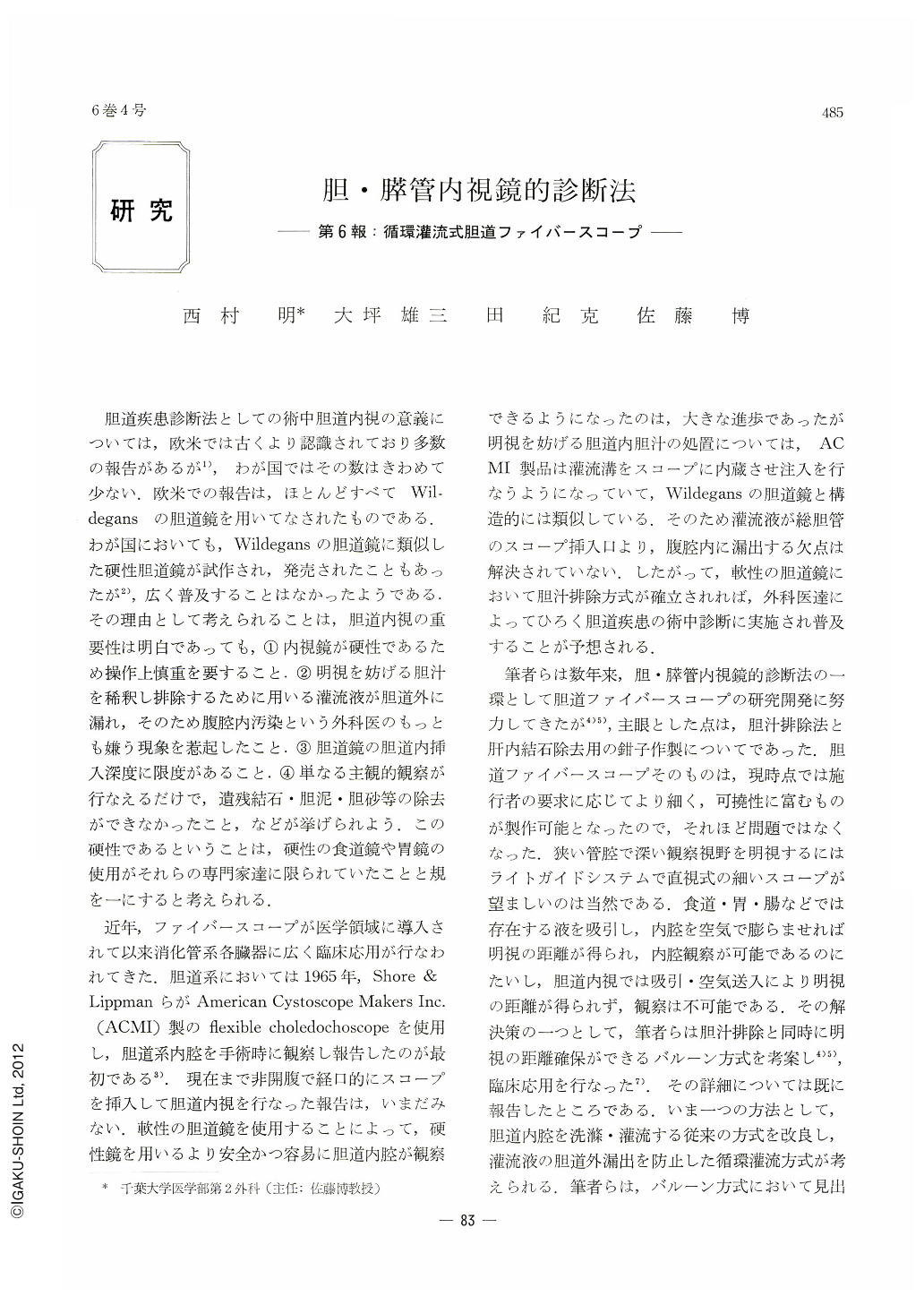
胆道疾患診断法としての術中胆道内視の意義については,欧米では古くより認識されており多数の報告があるが1),わが国ではその数はきわめて少ない.欧米での報告は,ほとんどすべてWildegansの胆道鏡を用いてなされたものである.わが国においても,Wildegansの胆道鏡に類似した硬性胆道鏡が試作され,発売されたこともあったが2),広く普及することはなかったようである.その理由として考えられることは,胆道内視の重要性は明白であっても,①内視鏡が硬性であるため操作上慎重を要すること.②明視を妨げる胆汁を稀釈し排除するために用いる灌流液が胆道外に漏れ,そのため腹腔内汚染という外科医のもっとも嫌う現象を惹起したこと.③胆道鏡の胆道内挿入深度に限度があること.④単なる主観的観察が行なえるだけで,遺残結石・胆泥・胆砂等の除去ができなかったこと,などが挙げられよう.この硬性であるということは,硬性の食道鏡や胃鏡の使用がそれらの専門家達に限られていたことと規を一にすると考えられる.
近年,ファイバースコープが医学領域に導入されて以来消化管系各臓器に広く臨床応用が行なわれてきた.胆道系においては1965年,Shore & LippmanらがAmerican Cystoscope Makers Inc.(ACMI)製のfiexible choledochoscopeを使用し,胆道系内腔を手術時に観察し報告したのが最初である3).現在まで非開腹で経口的にスコープを挿入して胆道内視を行なった報告は,いまだみない.軟性の胆道鏡を使用することによって,硬性鏡を用いるより安全かつ容易に胆道内腔が観察できるようになったのは,大きな進歩であったが明視を妨げる胆道内胆汁の処置については,ACMI製品は灌流溝をスコープに内蔵させ注入を行なうようになっていて,Wildegansの胆道鏡と構造的には類似している.そのため灌流液が総胆管のスコープ挿入口より,腹腔内に漏出する欠点は解決されていない.したがって,軟性の胆道鏡において胆汁排除方式が確立されれば,外科医達によってひろく胆道疾患の術中診断に実施され普及することが予想される.
As a method of endoscopy of the biliary tract in the midst of operation, the authors in the past exploited a choledochofiberscope with a balloon equipped at its distal tip, and have published the results of its clinical applications. In this paper is introduced a far improved choledochofiberscope manufactured for trial (CHF N0. 6) employing irrigation tube. Its characteristics, setup and directions for its use are described.
Although the new fiiberscope resorts to irrigation fluid the same as with irrigation methods hithereto used, an irrigation tube similar to tracheal tube is now employed to prevent fluid from leaking into the abdominal cavity so that dilatation of the biliary tract is all the more ensured. The fluid flowing within a irrigation circuit does not pollute the field of operation, and biliary sands and mud as well as pus within the biliary tract are washed away and discharged. Irrigation fluid is utilized as well for cytological diagnosis and bacilloscopy.
Directions for its use are simple and the procedure does not take much time. Less than 1,000 ml of fluid is sufficient for observation of the biliary tract lumen alone. There is no surface glare so harassing to the examiner in the baloon-tip method, and since it is not a contact view, wider field of observation is assured. Besides, the biliary tract mucosa as it is unstained by the bile is to be observed.
The authors now employ this method by preference in the observation of the lumen of the biliary tract. When co-employed with choledochography during operation, this method is assuredly a very eflicient procedure in the diagnosis and differential diagnosis in diseases of the liver and biliary tract.
Endoscopic pictures of several representative cases of inflammations, gallstones and tumors of the biliary tract as visualized by this method are also presented. Stress is laid on the fact that in cases of tumor cytological diagnosis of fluid contents collected during the operation plays a very important role in the accurate diagnosis of a neoplasm.
Copyright © 1971, Igaku-Shoin Ltd. All rights reserved.