Japanese
English
- 有料閲覧
- Abstract 文献概要
- 1ページ目 Look Inside
はじめに
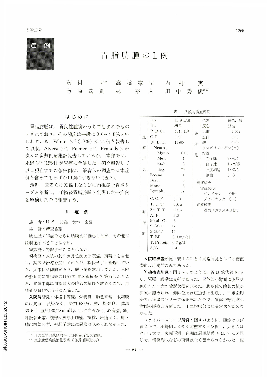
胃脂肪腫は,胃良性腫瘍のうちでもまれなものとされており,その頻度は一般に0.6~4.8%といわれている.Whiteら1)(1929)が14例を報告して以来,Alverzら2),Palmerら3),Peabodyらが次々に多数例を集計報告しているが,本邦では,水野ら5)(1954)が胃癌に合併した一例を報告して以来現在までの報告例は,筆者らの調査では本症例を含めてもわずか19例にすぎない(表2).
最近,筆者らはX線上ならびに内視鏡上胃ポリープと診断し,手術後胃脂肪腫と判明した一症例を経験したので報告する.
A 63-year-old woman complaining of headache and stiff shoulders was admitted to hospital on Dec. 1st, 1967 for the examination of the upper gastrointestinal tract.
At x-ray study free flow of contrast meal was seen through the esophagus into the stomach and duodenum with no evidence of hiatus hernia. A sharply circumscribed shadow defect the size of a hazel-nut was observed on the lesser curvature of the corpus. The stomach was of normal size, shape and location. Neither abnormal run of the mucosal folds nor mucosal convergence was seen. Gastric fiberscope also revealed a polypoirl lesion on the anterior wall near the lesser curvature at the level of the gastric angle. The surface of the tumor was smooth and of normal color. It was broad-based with no stalk noticiable. No bleeding was recognized on its surface or in the surrounding area. The mucosal folds were also normal. Its preoperative diagnosis was gastric polyp.
On Dec.11 of the same year total gastrectorny was done with Billroth II method. Pathologically it was lipoma in the submucosal layer consisting of mature fat cells encircled all around by collagen tissues.

Copyright © 1970, Igaku-Shoin Ltd. All rights reserved.


