- 有料閲覧
- 文献概要
- 1ページ目
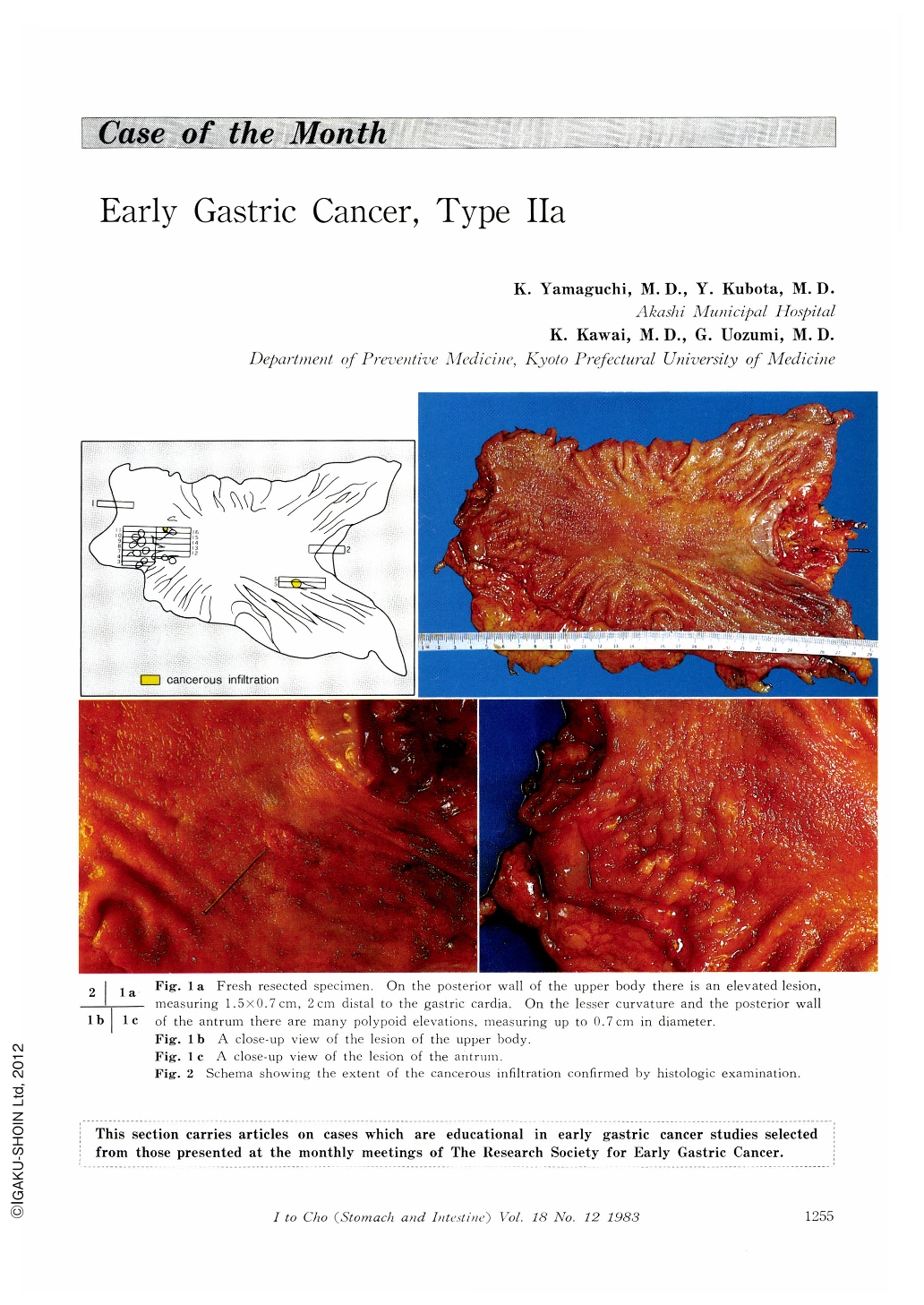
A 65 year-old house wife visited Akashi Municipal Hospital on January 25, 1983 with a chief complaint of nausea during meals which started in the last autumn. Her past history was unremarkable. Physical examination was normal. Laboratory tests were within normal limits. ECG showed mild myocardial ischemia. Upper gastrointestinal x-ray series were done on January 28, 1983 and revealed a small elevated lesion at the posterior wall of the upper gastric body and multiple polypoid lesions at the gastric antrum. The lesion at the upper gastric body was well-demarcated and its surface was slightly uneven with central depression. It was suspected of early gastric cancer, type Ⅱa. On the other hand, most of multiple polypoid lesions of the antrum had smooth surface with central depression, and were diagnosed as gastritis verrucosa.
Endoscopic examination with biopsy was performed on February 4, 1983. Dye-spraying method by indigocarmine was also done at the same time, and made it possible to obtain the better visualization of the elevated lesion at the posterior wall of the upper body. It was slightly discolored and lobular. Its surface was slightly uneven with a central depression. Early cancer, type Ⅱa, was suspected, and biopsy confirmed malignancy (Group Ⅴ). Another lesions at the antrum consisted of two different morphology on the endoscopic findings. One was reddish and smooth with or without central depression and the other, smaller and discolored without central depression. The former was diagnosed as gastritis verrucosa and the latter as intestinal metaplasia. Biopsy from one of the reddish polypoid lesions at the greater curvature revealed normal gastric mucosa (Group Ⅰ).

Copyright © 1983, Igaku-Shoin Ltd. All rights reserved.


