- 有料閲覧
- 文献概要
- 1ページ目
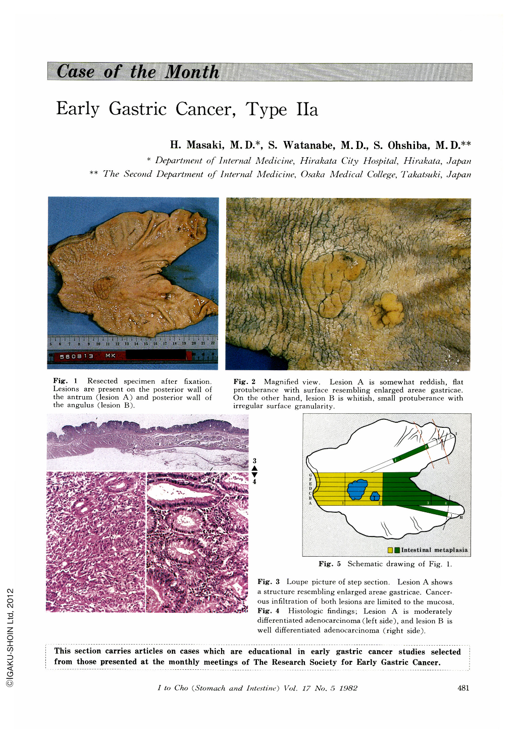
A 62-year-old man was admitted to Hirakata City Hospital in August, 1980. He had been treated in the out-patient clinic since 1979 because of diabetus mellitus. Upper G-Ⅰ x-ray and endoscopic examinations done in June, 1980 revealed abnormalities of the stomach. Family history was not contributory. Physical examination revealed no positive findings. Laboratory findings showed no abnormalities except for high fasting blood sugar level. Stool was negative for occult blood.
Endoscopic examination done on August 7, 1980 (Fig. 9, 10) demonstrated two lesions; the one, reddish, flat protuberant lesion with granular surface on the posterior wall of the antrum (lesion A); the other, uneven protuberant lesion with discolored surface on the posterior wall of the angulus (lesion B). The surface irregularity of the antral lesion was clearly demonstrated by direct spraying of Indigocarmin. It resembled the enlarged areae gastricae. Second x-ray examination was performed on August 10, 1980 (Fig. 5~8). Surface pattern of lesion A was delineated as the enlarged areae gastricae, and this lesion was well demarcated from the surrounding normal mucosa. Surface pattern of lesion B also revealed granularity.

Copyright © 1982, Igaku-Shoin Ltd. All rights reserved.


